Researchers at Ohio State University (OSU) in Columbus have developed a program to semiautomatically process MR images of the knee with the same -- and, in some cases, better -- accuracy as radiologists in one-fourth the time.
The software is designed to segment the meniscus in MR images to compare normal knees with knees from patients with moderate osteoarthritis. Having additional information about wear and tear on the meniscus could lead to the software's use as a biomarker in predicting which patients are at risk for developing osteoarthritis, the authors noted.
"From my past experience with different types of cancers, I know the signs of a disease are usually multidimensional and involve several things around the structure we are looking at," said co-developer Metin Gurcan, Ph.D., assistant professor in OSU's department of biomedical informatics. "That's why we started looking at the meniscus and began working to develop an algorithm to semiautomatically segment the meniscus from the MR images."
The research appears online and is scheduled for later print publication in the journal Osteoarthritis and Cartilage. The lead study author is Mark Swanson, a medical student at OSU.
Data collection
The researchers collected images and other data on 4,796 study participants in the Osteoarthritis Initiative, a national study of the disorder. They then randomly selected 24 images -- 10 images from patients with no symptoms of osteoarthritis and 14 from patients with confirmed cases of osteoarthritis.
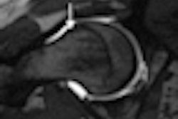
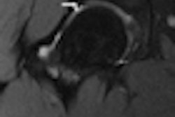
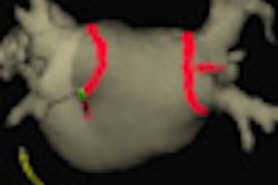
The program reads as many as two dozen MRI slices to designate and segment the 3D structure of the meniscus. As it moves through the images, the program compares the previous slice to the current slice, re-evaluates, and confirms its work.
Currently, the program requires that a radiologist scroll through the MR images manually, find the first slice that includes an image of the meniscus, and place a seed point within that area of the image. A second point must be placed on the meniscus in the last slice in which that part of the knee anatomy appears.
Once the segmentations are complete, clinicians can calculate volume, thickness, intensity, and any tears in the meniscus, which can be compared to calculations made with data from later images. If changes in the meniscus correlate with osteoarthritis symptoms, this part of the knee could become a target for preventing and treating the disorder.
Fast and accurate
The semiautomated segmentation method produced results similar to those of trained radiologists, with an average similarity index of more than 0.80 (a reading of 1.0 equals complete equivalence) for normal participants and 0.75, 0.67, and 0.64 for participants with confirmed knee osteoarthritis, according to the researchers.
"When we look at several readers' consistency and the computer's accuracy, the computer's accuracy was comparable to those of several readers," Gurcan said. In addition, the program took three to four minutes to complete its task, while it took the human readers 17 to 20 minutes.
The researchers currently are testing the software on a larger database to ensure its efficacy. "This process currently requires human interaction," Gurcan said. "You need to mark a seed point where the meniscus starts and where it ends. We are about to finalize another algorithm that will not require any human interaction at all. We will just give the images to the computer, which will completely automate the process."
Radiologist's tool
The long-range plan is to eventually make the software freely available to researchers around the world.
Even with the increased accuracy and faster processing of MR images, Gurcan emphasized that the process will not replace human radiologists.
"Over the years, I have encountered that question a lot," he said. "Everything we develop is just a tool that we provide to the radiologist, so we will never be able to replace the human decision at the end. What we are doing is trying to save them time and trying to increase their accuracy."
The research is funded through a grant from the U.S. National Institutes of Health (NIH).
To see a demonstration of the software, click here.
By Wayne Forrest
AuntMinnie.com staff writer
February 1, 2010
Related Reading
Sodium MRI at 3 tesla detects early signs of osteoarthritis, February 5, 2009
CAD shows potential for knee MR studies, May 26, 2008
U.K. study finds direct access to knee MRI is cost-effective, April 3, 2008
AAOS study: MRI overused for knee osteoarthritis, March 6, 2008
Presence of osteoarthritis influences bone bruise prognosis, August 22, 2007
Copyright © 2010 AuntMinnie.com