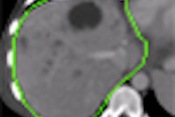
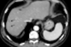
Differences with even modest 3D JPEG 2000 compression ratios can be visually perceived on thin-slice CT images of the chest and abdomen-pelvis, but it's unclear if these changes degrade diagnostic value, according to a multicenter study published online in the Journal of Digital Imaging.
Led by Dr. Bradley Erickson, Ph.D., of the Mayo Clinic in Rochester, MN, the research team sought to the determine the level at which 3D JPEG 2000 compression of these images becomes visually perceptible. They also wanted to see if residents in training and nonphysicians were substantially different from experienced radiologists in their perception of compression-related charges (JDI, July 15, 2009).
The researchers used 80 MDCT 3D datasets produced on systems from Siemens Healthcare of Malvern, PA. The datasets included 0.625-mm- to 1-mm-thick slices of standard chest, abdomen, and pelvis studies, clipped to 12 bits.
The studies had to be clipped to 12 bits to accommodate the compression algorithm (Kakadu Software) used in the study, which could handle only 8- and 12-bit grayscale images. The software was then used to compress and decompress the images to create four image sets: lossless; 1.5 bits per pixel (bpp) (8:1); 1 bpp (12:1); and 0.75 bpp (16:1). The average computation time to compress and decompress the studies was 30 minutes per volume on a 2-GHz-powered desktop personal computer.
The researchers then randomly selected slices from each examination for use during four observer sessions. In the sessions, the original and four compressed images were presented to readers in random sequences, according to the authors. Each sequence utilized only one occurrence of each image.
Observers used a special-purpose viewer application that showed images in a flicker mode, in which observers rapidly toggled between the original image and a compressed version to decide if they could detect any differences. Images were displayed on a Coronis color 3-megapixel LCD display (Barco, Kortrijk, Belgium) that was calibrated to the DICOM grayscale standard display function.
Users could select from four standard presentation window width and level settings, including lung, abdomen, bone, and liver. Image sizes were fixed at a 1:1 zoom to avoid possible interpolation effects, according to the researchers.
Three sites participated in the study and included six staff radiologists, four radiology residents, and six others with doctorates experienced in medical imaging.
The researchers found that overall, more differences were detected as compression levels increased (x² = 14,281.97, p < 0.0001), with 77.46% of observers detecting differences at 8:1, 94.75% at 12:1, and 98.59% at 16:1 compression levels. They also found 2.44% false-positive differences detected with the lossless images.
However, differences with even mild compression ratios were perceptible.
"We recognize that the diagnostic task is different, and in most cases, the diagnosis can be made with mild to even substantial visual differences," the authors wrote. "However, we feel that to be conservative, a visual difference should not be considered acceptable for any medical diagnosis that might be encountered. Other studies where specific diagnoses are considered cannot address the question of whether all diagnoses are unaffected."
No statistically significant differences were experienced overall as a function of body part imaged (x² = 0.150, p > 0.05). Of the trials with chest images, 67.82% had detected differences versus 68.79% of sessions with abdomen-pelvis images.
Statistically significant differences were noted, however, for observer experience (x² = 82.12, p < 0.0001). Overall, residents (71.91%) and those with doctorates (69.95%) detected more differences than the staff radiologists (64.70%) at all compression levels, according to the researchers.
Experienced radiologists appear to be less sensitive to compression-related changes than other observers.
"We found that the staff radiologists were consistently the least sensitive to compression artifacts," the authors wrote. "This may reflect the fact that staff was more comfortable that the images contained the information they needed even if slight changes were present."
In other findings, observers quickly found that liver settings were the most sensitive to compression-related settings, and reviewed most images with that setting, the researchers found. A significant difference (x² = 20.64, p < 0.0001) was also noted as a function of site.
While perceptible differences were found between lossy-compressed images and lossless images, that may not necessarily mean that important information was lost due to compression, the authors noted.
"Nevertheless, it appears that one cannot apply 3D JPEG 2000 compression to thin-slice CT studies at 8:1 or higher without considering possible effects on diagnosis," they wrote.
By Erik L. Ridley
AuntMinnie.com staff writer
August 19, 2009
Related Reading
German conference yields data compression recommendations, March 30, 2009
JPEG 2000 compression artifacts more prevalent in thin-section CT images, August 13, 2008
Lossy compression perceptible at even low levels, May 15, 2008
Lossy compression performs well in abdominal CT images, January 13, 2008
Canadian research finds no loss with lossy compression, August 14, 2007
Copyright © 2009 AuntMinnie.com
