Whole-body high-field MRI yields diagnostically accurate images of human fetuses, according to a study published online August 6 in the Lancet. As a minimally invasive postmortem exam, a whole-body high-field MRI exam may be more acceptable than a conventional autopsy to parents, who are often reluctant to approve this invasive procedure.
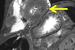
Researchers from the Centre for Cardiovascular Imaging at the University College London Institute of Child Health and Great Ormond Street Hospital for Children in the U.K. compared images from whole-body MRI at 1.5 tesla and 9.4 tesla using 3D T2-weighted fast spin-echo sequences. Both types of MRI exams were performed on 18 fetuses less than 22 gestational weeks of age, followed by a conventional autopsy.
Images from each MRI exam were interpreted in a blinded manner and compared with the findings of the autopsy. Tissue contrast of 14 different regions was compared for each set of MRI images in a random order, and image quality was scored on a four-point scale for diagnostic accuracy.
Spatial resolution, tissue contrast, and image quality of all organ systems were much better with high-field MRI than with conventional MRI, lead author Dr. Sudhin Thayyil and colleagues reported. The exams produced by high-field MRI detected all structural abnormalities that were identified by conventional autopsy, and with some cases, the high-field MRI images provided more detailed information than that discovered from autopsy.
Images from the conventional MRI exams were not diagnostically useful in 78% of the cases.
The researchers suggested that if the option of a noninvasive postmortem clinical evaluation were offered in lieu of a conventional autopsy, more parents might give their permission for such a procedure, restoring postmortem research in the U.K. Postmortem research of fetuses has been declining in the country for the past decade, according to Thayyil.
Copyright © 2009 AuntMinnie.com