A study from Oregon Health & Science University (OHSU) in Portland has concluded that 3-tesla MRI is superior to 1.5-tesla MRI in detecting and accurately characterizing structural brain abnormalities in a group of patients undergoing whole-brain epilepsy evaluation.
Researchers also found that 3-tesla MRI outperformed 1.5-tesla MRI in a direct comparison of lesion characterization, tissue contrast, and imaging artifacts. Three-tesla MRI correctly identified lesions in 88% of the cases analyzed, compared with 74% accurate identification for 1.5-tesla MRI.
The retrospective study, published in the American Journal of Roentgenology (September 2008, Vol. 191:3, pp. 890-895), reviewed 50 sets of MR images of 25 patients who underwent both 1.5- and 3-tesla brain imaging with a dedicated epilepsy protocol. Both protocols consisted of one 3D and three 2D sequences.
A six-channel sensitivity encoding head coil was used on both clinical 3-tesla units (Achieva, Philips Healthcare, Andover, MA) for all whole-brain epilepsy imaging. The 1.5-tesla MRI units (Signa Horizon and Signa LX, GE Healthcare, Chalfont St. Giles, U.K.) had a transmit-receive single-channel head coil for whole-brain imaging.
Patient sample
The study included 13 pediatric and 12 adult patients, ranging from 10 months to 70 years of age (mean age, 24 years). The mean time between 1.5- and 3-tesla MRI was 1.3 years, ranging from two months to five years. The study included all patients who received both 1.5- and 3-tesla whole-brain MRI at the Oregon facility between January 2000 and December 2005.
Four experienced neuroradiologists independently reviewed both 1.5- and 3-tesla MR images from the 25 patients. They rated both sets of images separately for lesion conspicuity, normal tissue contrast between gray and white matter, technical artifacts resulting in image degradation, and artifacts related to patient motion.
For each of the four quality parameters assessed, mean composite scores were higher for 3-tesla MRI than for 1.5-tesla MRI.
The readers' interpretations found statistically significant differences in three of four categories. In lesion conspicuity, 3-tesla MRI had a mean score of 2.95, compared with 2.29 for 1.5-tesla MRI. Three-tesla MRI also was significantly better than 1.5-tesla MRI in tissue contrast -- 2.73 versus 2.24, respectively. In technical artifacts, the readers rated 3-tesla MRI at 3.16, compared with 2.46 for 1.5-tesla MRI.
Lesion identification
In addition, 3-tesla MRI correctly identified structural lesions in 65 of 74 cases (88%), compared with 55 of 74 cases (74%) for 1.5-tesla MRI.
 |
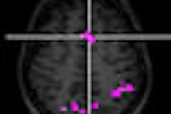
| Seventeen-year-old girl with intractable nocturnal seizures since the age of two. Above, coronal fluid-attenuated inversion recovery (FLAIR) 1.5-tesla MR image showing questionable curvilinear focus of juxtacortical high signal intensity (arrows) in left occipital lobe. Abnormal signal intensity was missed at first review of images. Below, coronal FLAIR 3-tesla MR image showing curvilinear band of high signal intensity (arrows) in left occipital juxtacortical white matter without apparent mass effect. Focus was surgically resected and histologic finding was focal cortical dysplasia with balloon cell features. |
 |
| Images courtesy of the American Journal of Roentgenology and Oregon Health & Science University; from Phal P, Usmanov A, Nesbit G, et al. Qualitative comparison of 3-T and 1.5-T MRI in the evaluation of epilepsy. AJR. 2008;191:890-895. Figure 1. |
The analysis also found that imaging artifacts were "less troublesome" at 3 tesla than at 1.5 tesla. "Although a trend toward greater motion artifacts was seen at 3-tesla, this difference was not statistically significant," the authors wrote. The four readers rated motion-related artifacts on 3-tesla MRI as 2.88, compared with 2.45 for 1.5-tesla MRI.
The researchers, led by co-author Dr. Pramit Phal in OHSU's division of neuroradiology, noted that the results of their study "support the clinical supposition that use of 3-tesla MRI increases the rates of lesion detection and accurate characterization of lesions. MRI at 3-tesla also yields better contrast resolution of the gray-white matter junction, a finding particularly relevant for detection of subtle focal dysplasia of the cortex."
Structural abnormalities
According to Phal and colleagues, the findings "imply that a 3-tesla MRI examination is 2.57 as likely as a 1.5-tesla examination to depict a structural abnormality and that correct characterization of the abnormality is 2.66 times as likely on 3-tesla studies as it is on 1.5-tesla studies, presumably contributing to a more accurate diagnosis."
The authors also noted several possible limitations of the findings. "The retrospective nature of the review, the indications for a second MRI examination at 3 tesla, and the need to exclude patients without directly comparable sequences may have introduced selection bias," they wrote.
Because the retrospective study could not control for the timing of the 1.5- and 3-tesla examinations, differences in visualization of certain conditions that change over time, such as tumor growth and conspicuity changes related to interim developmental myelination, may have influenced results.
By Wayne Forrest
AuntMinnie.com staff writer
October 1, 2008
Related Reading
PET/CT beats 3T MRI in whole-body primary tumor staging, March 10, 2008
3-tesla MRI sheds light on shading sign in endometriosis, June 8, 2007
3-tesla MRI goes mainstream with powerful new applications, March 1, 2007
Pilot study shows stereotactic radiosurgery safely treats epileptic seizures, December 6, 2006
Low-field MR with the right coil tops 3-tesla MR for prostate cancer imaging, December 15, 2005
Copyright © 2008 AuntMinnie.com