Computer-aided detection (CAD) technology is promising for the automated detection of meniscal tears, according to research presented at the recent 2008 Society for Imaging Informatics in Medicine (SIIM) meeting.
"We believe that the performance of the research CAD application was comparable to that of board-certified specialty-trained radiologists for the automated detection of meniscal tears," said Dr. Nabile Safdar of the University of Maryland School of Medicine in Baltimore. He presented the research during a scientific session at the conference in Seattle.
CAD has gained acceptance for certain image interpretation tasks, such as improved lesion detection, Safdar said. It may also improve reader efficiency depending on the application.
The researchers sought to evaluate the use of a homegrown CAD application for automatic detection of meniscal tears and articular cartilage injuries of the knee. These tears are common in young athletes and the aging population, requiring accurate diagnosis and, if appropriate, surgical intervention.
The study team retrospectively searched a pre-existing report repository for knee MRI cases between October 2003 and July 2006 using the following search terms: meniscal tear, horn, knee, and arthroscopy. All ages and sexes were included.
A total of 40 cases were collected, 17 of which had meniscal tears. Arthroscopic results were gathered from the electronic medical records to serve as a reference standard.
An additional 60 cases were randomly identified from a national osteoarthritis database, and two board-certified radiologists with musculoskeletal fellowship training provided interpretations of these cases to provide the reference standard.
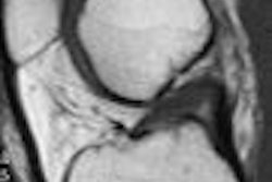
MRI scans from both groups were performed using either an Avanto or Espree 1.5-tesla MRI scanner (Siemens Healthcare, Malvern, PA). The osteoarthritis database cases utilized a double-echo steady-state (DESS) MR sequence, while the locally acquired images were T1-weighted sequences with a 512 x 512 matrix, Safdar said.
The two board-certified radiologists were asked to evaluate the 40 cases generated locally for the presence of meniscal tears without the assistance of CAD. For the sake of accuracy, menisci were divided by the researchers into four groups.
Nonphysician researchers then applied the CAD algorithm to the same dataset. The accuracies of both radiologists were calculated and compared with the CAD results. The 60 cases with the articular cartilage abnormalities were interpreted and evaluated in a similar way, according to Safdar.
The radiologists turned in a sensitivity of approximately 84% and specificity of 77% for evaluating meniscal tears, while the CAD algorithm had a sensitivity of approximately 75% and a sensitivity of 81%. There was no significant difference in overall accuracy between the radiologists and the CAD system, Safdar said.
CAD did not perform as well in evaluating articular cartilage, however. Radiologists had a sensitivity of approximately 92% and a specificity of 96%, compared with sensitivity of approximately 85% and a specificity of 72% for the CAD system. There was a significant difference in overall accuracy between the radiologists and the CAD for the articular cartilage cases (p < 0.05).
"We do think we need to improve the accuracy of the CAD algorithm in the evaluation of articular cartilage injuries," Safdar said.
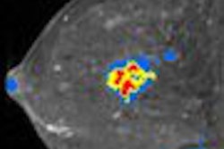
For evaluating meniscal cartilage, the CAD application performed a two-stage process. The first stage consisted of region of interest (ROI) selection, automatic slice selection, binarization, and the enforcing of shape constraints, according to Safdar. The second stage included the scoring of the slices for potential tears and the generation of the final recommendation of either a torn or normal meniscus.
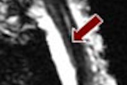
To evaluate the articular cartilage, the CAD application employed a 2D active shape model (ASM) to model the bone-cartilage interface on all slices of the DESS MR sequence. It then measured the cartilage thickness from the surface of the bone and identified the regions of abnormal thinness and focal/degenerative lesions, according to the researchers.
Safdar noted that the researchers were pleased that although the application wasn't as accurate in articular cartilage abnormalities, there was higher specificity or fewer false-positive markings than some other CAD applications, adding that "this is a very different type of pathology."
Future research will include analysis of CAD's effect on interpretation time and reader confidence, as well as a prospective study comparing radiologists with and without CAD, Safdar said.
By Erik L. Ridley
AuntMinnie.com staff writer
May 26, 2008
Related Reading
VC CAD finds polyps in prepped or unprepped patients, May 6, 2008
False-positive CAD marks don't hinder VC readers' accuracy, April 21, 2008
Reimbursement crunch throttles breast MRI CAD users, April 8, 2008
Criteria determine CAD mark sensitivity, March 20, 2008
CAD provides mixed benefits for DR lung exams, March 8, 2008
Copyright © 2008 AuntMinnie.com