Automated fetal biometry measurements compare well with measurements performed by sonographers, according to research presented at the recent American Institute of Ultrasound in Medicine (AIUM) annual meeting.
"Automation of full ultrasound measurement has great potential for improving productivity and patient throughput, enhancing accuracy and consistency of measurements, and reducing the risk of repetitive stress injury," said Dr. Ivica Zalud of the University of Hawaii in Honolulu. He presented the research findings during a scientific session at the San Diego show.
Automatic measurements can reduce keystrokes and potentially reduce repetitive stress injuries, Zalud said. In addition, they have the potential to improve everyday workflow to increase patient throughput and productivity, and also decrease interobserver variability.
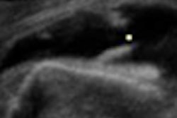
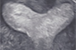
The study team sought to compare the performance of Auto OB software (Siemens Medical Solutions, Malvern, PA) and sonographers in measuring biparietal diameter, head circumference, abdominal circumference, and femur length. They performed two sets of experiments involving five experienced sonographers with at least five years of experience; the first set was designed to assess the performance of the automated approach relative to the expert users in 250 images for each user.
The researchers set a target performance for the automated system for an average error rate of less than 3% in 80% of the cases. In the second set, the researchers sought to compare the automated system with a ground truth generated by five expert users using a set of 10 images per anatomy.
"Given that all users made measurements on the same set of 10 images per anatomy, we have five different measurements per image, which allowed us to compute a common ground truth by taking the average of those measurements," Zalud said.
The range of sonographer errors was 0.01% to 6.36%, while the automated system had a range of 0.07% to 3%. The automated system had an average error of 1.89% in 80% of the cases for the four measurements, significantly better than the 3% target error rate, he said. The Pearson correlation coefficient was 0.998.
In the second exam set, the automated system had a mean error of 1.8%, while the users had a mean error of 1%. That difference was not statistically significant, according to Zalud.
"This error difference typically is an error of assessment of ± 1 to 3 days in the worst-case scenario at the end of the pregnancy when dispersion of measurements is the highest," he said.
The researchers noted that the Pearson coefficient correlation was lowest for biparietal diameter (r = 0.996) and highest for abdominal circumference (r = 0.999).
"Measurement errors produced by Auto OB translated into very small errors regarding (gestational age) of the fetuses," Zalud said.
An error rate of 1.8% represents a deviation of less than two days for 20 weeks of gestational age (GA), less than four days for 30 weeks of GA, and less than seven days for 40 weeks of GA, he said.
"Remember, we still teach our residents that our error in measurements in the third trimester could be as much as three weeks," Zalud said.
By Erik L. Ridley
AuntMinnie.com staff writer
March 24, 2008
Related Reading
GA-oriented morphology boosts fetal US malformation workup, August 9, 2007
Fetal entertainment ultrasounds draw patient interest, March 20, 2007
Early prenatal ultrasound inadequate for detecting congenital heart malformations, June 30, 2006
Empty renal fossa on prenatal ultrasound indicates renal ectopia or agenesis, August 17, 2005
Fetal ear length may indicate chromosomal abnormality, April 11, 2002
Copyright © 2008 AuntMinnie.com


