VIENNA - Following last week's release of a major study showing that flat colorectal lesions are more common and more dangerous than initially thought, the pressure is on for colon screening modalities, particularly virtual colonoscopy (VC or CT colonography [CTC]), to show they can routinely detect lesions characterized by flat or concave morphology.
If flat lesions are a challenge for conventional colonoscopy, they are considered even harder to find at VC due to their radiologic subtlety. If radiologists are to maintain a high sensitivity level for detecting these lesions, radiologists reading VC may need help.
However, in light of the U.S. Food and Drug Administration's virtual "freeze" on new 510(k) approvals for colon CAD systems in the past several years (pending investigation into several areas of concern, according to the FDA), the worry among U.S.-based VC practitioners is that help won't come soon enough.
But study results are encouraging. In two studies presented Sunday at the 2008 European Congress of Radiology, investigators specifically tested colon computer-aided detection (CAD) schemes for their ability to find flat lesions. In both, the sensitivity was reassuringly high, though questions remain about the effect of false-positive CAD detections on reader performance and variations in flat polyp morphology.
The flat-lesion bombshell that has grabbed the attention of researchers worldwide appeared March 5 in the Journal of the American Medical Association. In the large, single-center study, nonpolypoid lesions were found in about one in 11 individuals undergoing elective optical colonoscopy.
Study authors Dr. Roy Soetikno et al from the Veterans Affairs Palo Alto Health Care System in Palo Alto, CA, examined 1,189 patients in a Veterans Affairs healthcare setting. The 170 nonpolypoid lesions they detected contained 15 cancers, nearly 10 times the rate (odds ratio, 9.78; 95% CI, 3.93-24.4) of polypoid lesions, irrespective of lesion size (JAMA, March 5, 2008, Vol. 299:9, pp. 1027-1035).
Sphericity settings affect flat lesion detection
Some studies suggest that as many as one-fifth of cancers may have flat morphology, a lesion type that is difficult to detect at VC, said Dr. Stuart Taylor of University College London in the U.K. in his ECR presentation.
In the past several years, colon CAD software has become increasingly robust at finding polypoid lesions; however, its performance in flat lesions is poorly understood, he said.
"We also know that most CTC reader errors are perceptual errors where people don't see the lesions; therefore, CAD may have an important role," Taylor said. "Combining the high clinical importance and radiological subtlety of flat lesions makes an ideal target for CAD."
To assess CAD's peformance in early-stage, morphologically flat lesions, researchers from University Hospital London, with colleagues from Beijing Friendship Hospital in Beijing and the National Cancer Center in Tokyo, performed a retrospective study of 30 symptomatic patients with stage T1 lesions classified as nonpolypoidal (Paris classification stage 0-II). The cases were staged at CTC following endoscopic detection.
All CTC exams were performed on a 64-slice scanner (Aquilion 64, Toshiba Medical Systems, Tokyo) using 120 kVp, 200-400 mAs, 0.5-mm collimation, and supine image acquisition following the adminsitration of intravenous oral contrast.
A single experienced radiologist applied CAD software (ColonCAD API 4.0, Medicsight, London) integrated into the Vitrea workstation to the cases.
"The particular CAD we used has a so-called sphericity filter, and what it does is analyze every voxel on the candidate surface to determine whether it or its neighbors form part of a theoretical perfect sphere," Taylor explained. When analyzing data the sphericity filter can be aimed at findings with more or less perfect sphericity in settings ranging from 0 to 1, he said, as follows:
- Sphericity score 1: voxels part of "perfect sphere"
- Sphericity score 0: voxels form part of an increasingly less perfect sphere
- Sphericity score 0.75: current system default for polypoidal lesions
The researchers applied the CAD application at all three sphericity setttings (0, 0.75, 1) for each dataset. A CAD mark directly overlapping a flat lesion was deemed a true positive, and further subclassified as focal or nonfocal depending on whether it was placed directly over a flat lesion or a nondefinitive region of the lesion. All CAD marks not located over a lesion were deemed false positives, Taylor said.
 |
| Each CAD mark over a flat lesion was deemed a true positive, which was further classified as focal or nonfocal depending on the location of the mark over the lesion. Lesions with a CAD mark that was located but not centered on the polyp were deemed nonfocal. |
The CTC results yielded a total of 24 pure type II lesions among the 30 patients, located in the rectum (n = 4), the sigmoid colon (n = 6), the descending colon (n = 4), the transverse colon (n = 6), the ascending colon (n = 3), and the cecum (n = 1).
 |
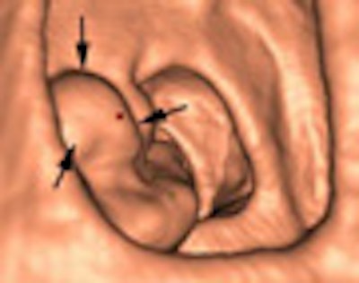
| The Paris classification system macroscopically defines variations in polyps with flat morphology, defined as lesions with more than twice the length as height. Type IIa is the most commonly detected type and the easiest type for CAD to find. All images courtesy of Dr. Stuart Taylor. |
The mean lesion size was 25 mm (range, 7 mm to 60 mm). Twenty of 24 cancers were seen on supine and prone datasets; three were visible on supine only, and one was seen on prone only.
The results showed varying sensitivity by flat lesion type and sensitivity for CAD, which detected 20 of 24 (83.3%), 17 (70.8%), and 13 (54.1%) of the flat lesions at sphericity settings of 0, 0.75, and 1, respectively.
 |
| The results show varying sensitivity by flat lesion type and sensitivity for CAD. Sensitivity was highest (83.3%) when sphericity was set at 0. However, this setting significantly increased the number of false positives. The mean number of false positives per patient was 36.5, 21.1, and 9.5 at sphericity settings of 0, 0.75, and 1, respectively. |
"As we've seen in other presentations, CAD tends to hit these lesions quite a lot," Taylor said. In his group's study, "there are at least two CAD marks for every (detected) flat lesion," he said.
However, the lower the sphericity setting, the greater number of false positives generated, Taylor added. The mean number of false-positives per patient was 36.5, 21.1, and 9.5 at sphericity settings of 0, 0.75, and 1, respectively.
Sensitivity was highest (83.3%) when sphericity was set to 0, he said. However, this setting significantly increased the incidence of false positives. The mean number of false positives per patient was 36.5, 21.1, and 9.5 at sphericity settings of 0, 0.75, and 1, respectively.
 |
| The number of false-positive detections increased when sphericity settings were decreased. Chart shows the causes of false-positive findings for each setting and lesion type. Most occurred in normal mucosa. |
"Most flat polyps are detectable at CAD," Taylor said. "You can maximize your sensitivity by use of a filter. Type IIa is the best detected perhaps due to the focal protuberances."
There is a trade-off between sensitivity and the false-positive rate, and one can argue over which is better: CAD with 25 false posiives that detects a flat cancer, or CAD with five false positives that misses it. Reader studies will be needed to confirm the results, he said.
High sensitivity with fewer false positives
In another ECR presentation, Marcos Salganicoff, Ph.D., from Siemens Medical Solutions of Malvern, PA, discussed research on the firm's investigational colon CAD application.
The study sought to analyze performance of the CAD algorithm in a standalone setting, and on various types of polyp morphologies: flat, sessile, and polypoid. The research is "driven by the fact that our CTC CAD system users expect our system to detect the entire spectrum of lesions in routine cliical use, including lesions that are more challenging not only for the human reader, but also for the CAD system," Salganicoff said.
In all, 418 CTC cases were acquired from 10 sites in the U.S. and Europe. The investigators used a variety of standard kernels and acquistion parameters (all 1-mm to 2-mm collimation) on various scanners; all subjects had been cleansed cathartically, he said. The dataset contained 174 polyps ranging from 6 mm to 25 mm in diameter. In all 208 cases were used to "train" the CAD system, while the remaining 210 pateints consituted the test group.
In the test group, 99 cases were positive,and included 65 medium-sized (6 mm to 9 mm) lesions, and 38 large-sized (9.9 mm to 24.9 mm) lesions. The sensitivity for sessile and pedunculated polyps was 100% for both medium- (25 of 25) and large-sized (16 of 16) polyps. As for flat polyps, CAD sensitivity was 93.8% (15 of 16) for medium-sized lesions and 92.3% (12 of 13) for large-sized polyps, Salganicoff said.
The study did not utilize the Paris classification system. Per-polyp CAD sensitivity for shallow flat polyps, defined as those with a height of less than 3 mm, was 97.6% for medium-sized lesions and 96.5% for large-sized lesions, with an average of 2.7 and a median of two false positives per exam volume.
"With flat lesions, we have a drop in performance that is still quite good, and obviously the sample size is (small) for large flats, so it's difficult to conclude," Salganicoff said. The false-positive rate was quite good, considering the prevalence of polyps in the clinical setting, he added.
"We had a mean two (false positives) per volume so we expect about four to five (per patient) with prone and supine data combined," Salganicoff said. "There are no sphericity settings; the system has a preset threshold," he said. "We've demonstrated very high sensitivity with a very good false-positive rate for all polyp morphologies, including flat polyps and shallow-flat polyps."
Limitations included the use of data from multiple centers, including some with less-than-optimal prep. Those cases that were not colonoscopy-confirmed were at least double-read, Salganicoff told AuntMinnie.com.
By Eric Barnes
AuntMinnie.com staff writer
March 9, 2008
Related Reading
Flat colorectal tumors relatively common in U.S. population, linked to cancer, March 5, 2007
American Cancer Society recognizes virtual colonoscopy screening benefit, March 5, 2008
Noncathartic bowel prep facilitates, complicates virtual colonoscopy, June 15, 2007
New developments improve VC -- and colonoscopy, April 6, 2007
Prepless VC misses some diminutive flat lesions, January 17, 2006
Copyright © 2008 AuntMinnie.com