Japanese researchers have produced good results using a digital tomosynthesis technique for general radiography studies of the temporomandibular joint (TMJ). As part of its research, the group also developed a new reconstruction algorithm for digital tomo studies that they say reduces image artifacts in the 3D volumes generated by the digital tomo system they used.
The TMJ is a difficult area to image due to the complex anatomy of the joint. Conventional radiography suffers limitations due to the superimposition of anatomy and metallic restorations over regions of interest, the researchers wrote in a paper published this month in Dentomaxillofacial Radiology (December 2007, Vol. 36:8, pp. 514-521).
Clinicians are putting newer modalities such as MRI and CT (especially conebeam CT) to work in the TMJ, but these technologies also have disadvantages. MRI can be too expensive for many practices, while CT produces higher radiation doses than radiography.
The group sought to assess whether a relatively new radiography imaging technique, digital tomosynthesis, could address some of these drawbacks. Digital tomosynthesis involves the use of a specially outfitted digital radiography system that has an x-ray tube and digital detector that move in an arc around the region of interest.
The technique captures multiple tomographic slices that are then reconstructed into a single 3D image. Digital tomo advocates believe that the technology improves visualization of hard-to-see structures by reducing the interference of overlapping anatomy on a particular region of interest.
Researchers at Kitasato University in Sagamihara and Shinshu University in Asahi decided to explore the effectiveness of digital tomosynthesis for TMJ imaging using a digital x-ray system outfitted for the technique (Sonialvision Safire, Shimadzu Medical Systems, Kyoto, Japan). The study was supported by funding from Shimadzu.
In addition to the digital tomo unit, the researchers also wanted to assess the effectiveness of a new data reconstruction technique they felt could address some of the drawbacks of a commonly used 3D reconstruction algorithm, the Feldkamp 3D filtered back-projection (FBP) technique.
FBP has been used for digital tomo and conebeam CT reconstructions, but it can produce artifacts when objects are separated from the central plane to the far direction, the researchers wrote. They developed what they called a modified FBP algorithm that uses low-pass filtering along the sectional axis of Fourier space data that has undergone a 3D Fourier transform.
The researchers first conducted digital tomo studies on an anthropomorphic phantom to assess the effectiveness of the modified FBP algorithm compared to the older Feldkamp technique. They then conducted digital tomo studies with modified FBP reconstruction on patients, and compared the images to both panoramic radiography and conventional tomography.
In the phantom studies, the group created datasets with 90 images at a tomo angle of 40°. The digital x-ray system used imaging parameters of 80 kVp and 160 mA, with an exposure time of 200 msec per view. The plane locations were the center plane (0 mm) and an off-center plane 70 mm from the center plane.
The researchers found that the modified FBP algorithm produced digital tomo images that were sharper and had fewer artifacts than the conventional Feldkamp algorithm. The images reconstructed with the Feldkamp algorithm were "dominated by blurring artifacts," while the artifacts were mostly removed from the modified FBP images, resulting in higher spatial and contrast resolution.
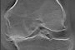
In the patient studies, the tomo technique offered improvements over both panoramic radiography and conventional tomography. "The use of digital linear tomosynthesis improved visualization of the underlying tissue detail by blurring the overlying structures," the researchers wrote. "This allowed better visualization of the tissue detail directly below the TMJ structures."
The effectiveness of digital tomosynthesis is strongly affected by scan parameters, including tomographic angle, number of views, and section thickness, the group concluded. For TMJ imaging in particular, a large number of views are required due to the complexity of the anatomical structures in the area, they said.
However, digital tomosynthesis produces high-quality images, particularly with coronal views, and complete studies can be conducted in a few minutes. The researchers suggested that digital tomo be considered the technique of choice for investigating bony changes of the TMJ.
By Brian Casey
AuntMinnie.com staff writer
December 24, 2007
Related Reading
DR pumps new life into tomosynthesis-based radiography, October 29, 2007
Duke researchers combine DR tomosynthesis with 3D CAD, June 4, 2007
Defining a role for MRI in TMJ disorders, October 13, 2006
Copyright © 2007 AuntMinnie.com