PROVIDENCE, RI - As FDG-PET's role in monitoring tumor response to therapy gains popularity, questions have arisen about the reliability of various methods to determine regions of interest (ROI) to measure FDG uptake.
Researchers from the University of California, Los Angeles (UCLA) sought to shed some light on the topic in their study to determine the effects of ROI definition on quantitative measurements of tumor FDG uptake.
Vladimir Evilevitch from the department of molecular and medical pharmacology in UCLA's David Geffen School of Medicine presented the results of the study at the first annual joint conference of the Academy of Molecular Imaging (AMI) and Society of Molecular Imaging (SMI).
Researchers analyzed 41 patients with locally advanced, high-grade bone and soft-tissue sarcomas. Twenty-two patients were male. Patients had a mean age of 47 years and had received chemotherapy prior to surgery.
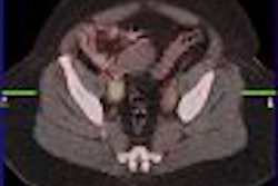
A baseline PET/CT scan was performed within four weeks of surgery, while a follow-up scan was done within three weeks before surgery. Patients received 0.21 millicuries (mCi) of FDG 60 minutes prior to imaging.
SUV measurements
To measure tumor FDG uptake, researchers used three factors: maximum FDG uptake (SUVmax) within the tumor; FDG uptake within a 1.5-cm ROI around the maximum tumor volume (SUVpeak); and mean uptake (SUVvolume) based on the volume of the whole metabolically active tumor.
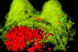
To determine SUVmax, researchers designated a rectangular ROI around the entire tumor in all axis slices to demonstrate focal FDG uptake. The pixel with the maximum FDG uptake in this region was highlighted. They then drew a circle with a diameter of 15 mm around the pixel to calculate the mean SUV within this ROI to use as the SUVpeak.
In addition, computer-generated isocontours were used to outline the metabolically active tumor volume, with 50% threshold for the maximum FDG uptake in the baseline scan. A manual correction was performed to exclude normal tissue with physiological FDG uptake. The mean SUV within this volume of interest also was determined.
The next step was to study two approaches to assess tumor response to pretreatment therapy by FDG-PET. In the first approach, researchers measured SUVmax, SUVpeak, and SUVvolume after the completion of therapy. In the second approach, the study determined changes in SUVmax, SUVpeak, and SUVvolume from the baseline to the post-treatment scan. The standard for tumor response was a patient's histopathological data.
Post-therapy response
As in previous studies, tumors were classified as responding when there was at least 90% tumor necrosis after therapy. In cases of no necrosis, tumors were classified as nonresponding.
"In the post-treatment scan, all three parameters -- SUVmax, SUVpeak, and SUVvolume -- were significantly lower in histopathological responders than in nonresponders," Evilevitch noted in his presentation. "Similarly, changes in FDG uptake from the baseline to the follow-up scan were significantly more pronounced in responders than nonresponders."
At the same time, the study also found that average SUVs within the responder and nonresponder groups varied substantially. For example, the average post-treatment SUV of the histopathology nonresponder group was 6.7 for SUVmax and only 2.4 for SUVvolume. The average post-treatment SUV in the responder group was 3.3 for SUVmax and 1.1 in SUVvolume.
In addition, relative changes in SUVs were similar in SUVmax, SUVpeak, and SUVvolume, as the average change in the responder group ranged from 62% for SUVpeak to 68% for SUVvolume. Relative changes in the nonresponder group ranged from 33% for SUVmax to 40% for SUVvolume. "Thus, relative changes were much less influenced by the way regions of interest were drawn," Evilevitch said.
ROC analysis
When the study reviewed results based on receiver operating characteristics (ROC), SUVmax, SUVpeak, and SUVvolume overlapped "to a great extent," Evilevitch added, "indicating that all three parameters provide a similar accuracy for assessment for histopathological response."
"However," he continued, "the threshold values for providing the highest sensitivity and specificity for differentiation of responders and nonresponders shows large variability (ranging from 1.3 for SUVvolume to 5.2 for SUVmax), again demonstrating that post-treatment SUVs are very sensitive to region of interest size."
From the study results, UCLA researchers concluded that SUVs of sarcomas after therapy are "highly dependent" on ROI definition, while treatment-induced changes in SUVs are only "mildly affected" by significant differences in ROI definition.
"This indicates that changes in SUVs represent more robust parameters for assessment of tumor response" in clinical trials, Evilevitch said.
By Wayne Forrest
AuntMinnie.com staff writer
September 11, 2007
Related Reading
FDG-PET can influence Alzheimer's, dementia treatment, September 6, 2007
FDG-PET could help unlock secrets of atherosclerotic plaque, August 2, 2007
SUV readings vary on different PET systems, study finds, July 25, 2007
PET changes oncology management, study shows, July 19, 2007
FDG uptake correlates with survival, biomarkers in NSCLC, June 19, 2007
Copyright © 2007 AuntMinnie.com