Scientists at the University of Alberta Cross Cancer Institute in Canada are constructing a system that couples a linear accelerator (linac) with an MR scanner that will provide real-time tracking of tumors and healthy tissues during irradiation. A prototype of the fixed system is expected to be completed by this December. The Alberta Cancer Board has received U.S. and international patents for the system, called Advanced Real-Time Adaptive RadioTherapy (ART).
Alberta Cancer Board researcher Gino Fallone, Ph.D., said this hybrid radiation system could overcome the problems of patient motion and poor tumor definition that currently plague x-ray and CT imaging.
"Our system will be beneficial for all solid cancers, but will especially benefit those cancers that are difficult to treat with today's radiation technology, such as liver, stomach, and pancreas, and significantly improve on other cancers where it is still difficult to administer sufficient radiation dose to obtain a good chance of cure, such as lung cancer, for example," said Fallone during a presentation at the 2007 American Association of Physicists in Medicine (AAPM) meeting in Minneapolis. Fallone designed the ART system along with Marco Carlone, Ph.D., and Brad Murray, Ph.D., also with the Alberta Cancer Board.
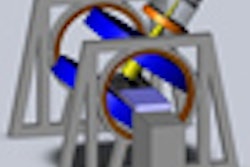
The linear accelerator and an MRI system are fixed with respect to each other, and rotate in unison around the patient, delivering treatments from all angles. Magnetic shielding prevents the MRI from disturbing the linac, and radiofrequency (RF) signal shielding, modifications to RF-signal triggering, and pulse shaping are used to minimize the linac's electromagnetic interference with the MRI.
 |
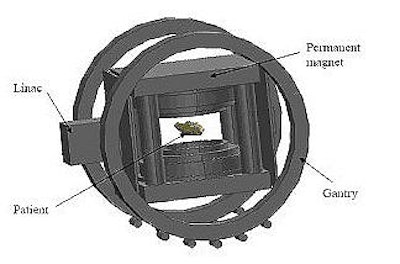
| Diagram of the prototype hybrid MRI-radiation therapy machine. Advantages include an unobstructed treatment beam and real-time imaging during radiation therapy. Image courtesy of Gino Fallone, Ph.D., Alberta Cancer Board. |
The biplanar, open MRI system has a 65-cm opening to allow the rotation of the shoulders within the bore, the authors stated in their AAPM abstract. This allows a patient's head to remain outside the machine for most procedures.
The system addresses the problem of organ motion by tracking the moving tumor in real time with MR imaging, and adjusting the radiation beam accordingly. Because the imaging and radiation beam are both fixed on the patient, the radiation beam remains "locked on" the tumor despite breathing or other organ movement. The inventors believe this will make it possible to reduce the treatment margin of error typically built into treatment plans to account for movement and other factors, thus reducing damage to surrounding healthy tissue.
Clinicians typically calculate beam intensity and location for treatment plans based on various imaging exams, including radiographs, CT, ultrasound, MRI, and PET. These complex calculations must also factor in treatment margins to account for organ movement, as well as body changes that may occur in the time between imaging, mapping and the radiation application.
Use of MRI real-time imaging coupled with linac tracking will provide higher-quality images of tumors and organs than x-rays and CT, and adds precise beam application, according to Fallone.
Fallone told AuntMinnie.com that his group had not done any animal experiments with the device, but do plan to start testing on human subjects in the near future. He declined to say whether the group is currently working with any vendors on this project.
By Kathlyn Stone
AuntMinnie.com contributing writer
September 14, 2007
Related Reading
Radiation oncology and PACS: ramping up for the digital age, August 9, 2007
Radiologists, clinicians perceive PACS success differently, June 27, 2007
SPECT/CT offers on-target diagnosis in noncancerous bone disease, March 23, 2007
Copyright © 2007 AuntMinnie.com