Researchers in Japan and the U.S. are producing promising new head CT images on a work-in-progress 256-slice scanner. A major advantage of so-called area detector scanning is the ability to acquire morphologic and functional images in a single scan, yielding a clutch of new diagnostic possibilities and time-saving exam protocols.
At the 2007 European Congress of Radiology (ECR) in Vienna, Dr. Kazuhiro Katada, chairman of the department of radiology at Fujita Health University School of Medicine in Japan, discussed his group's recent work with head and brain imaging at 256-slice CT. He followed up with additional information in a series of e-mails to AuntMinnie.com.
Katada and his colleagues are using Toshiba Medical Systems' investigational Aquilion 256 CT scanner for research at Fujita. A second scanner was installed in January at Johns Hopkins University in Baltimore for a six-month project to study cardiac and head imaging applications, Katada said.
The system uses the same detector module as Toshiba's 64-slice Aquilion, arranged in a 256 x 0.5-mm configuration, and generating 128 mm of coverage per rotation. There are 912 channels x 256 segments, with an element size of approximately 1 x 1 mm, which corresponds to a 0.5 mm (transverse) x 0.5-mm (longitudinal) beam width at the center of rotation. The gantry rotation time is 1 second and the data sampling rate is 900 views per second.
The slow rotation speed is due to limitations of the prototype, but will be substantially faster in the commercial model, Katada said. Toshiba said it anticipates U.S. Food and Drug Administration clearance and commercial release of the scanner within a year.
The 256-row area detector CT scanner enables scanning over a 128-mm range to be performed in a single rotation, without the need for helical scanning, Katada said. This will enable the acquisition of high-resolution perfusion data of the entire brain to be produced very quickly.
In his ECR presentation, Katada called head imaging "a really exciting area of research," owing to the 256-slice scanner's ability to produce high-resolution images in a very short time, with "positive effects on workflow."
 |
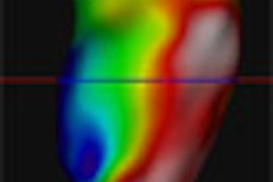
| Whole-brain CT perfusion at 256-slice CT enables the fast acquisition of high-resolution perfusion images. All images courtesy of Dr. Kazuhiro Katada. |
Head images can be acquired two ways on the 256-slice machine: in so-called "snapshot" scans or by sequential scanning in continuous acquisition mode, which can be used as a data source for whole-brain CT perfusion, subtraction CT angiography, dynamic multiplanar reconstructions (MPRs), and other uses, all in a single acquisition, Katada said.
The current generation of multislice scanners acquires only four slices at a time, while 256-detector-row CT can provide volumetric cine imaging and perfusion CT simultaneously in a wide craniocaudal coverage area, he said.
Another limitation of perfusion scanning with helical CT is that repeated scanning of the same anatomic region increases radiation exposure, Katada said. To address this problem, the Fujita researchers used a low-dose protocol of 80 kVp and 80 mAs, combined with intermittent scanning.
At these settings, the signal-to-noise ratio is poor due to photon starvation, "so we have employed image averaging," Katada said. "This is just like functional imaging in MRI, and it works perfectly."
 |
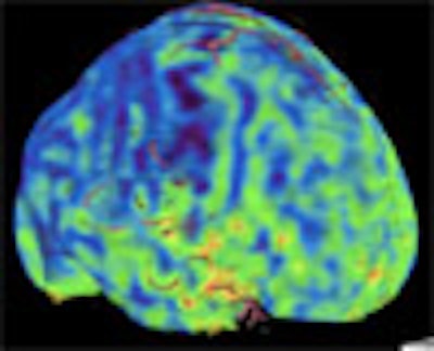
| In a patient with Moyamoya disease, time sequential averaging compensates for a lack of photon penetration in the low-dose protocol, improving the signal-to-noise ratio. |
As of this month, Katada and his colleagues have performed a total of 20 head scans using 0.5-mm slice thickness at 1-second intervals for the arterial phase, and 3-second intervals for the venous phase over 50 seconds (1-second rotation), for a radiation dose of 5-7 mSv, he explained.
Since the area detector permits perfusion data to be obtained as a single volume covering the entire brain, sagittal images, coronal images, and volume-rendered images, as well as standard axial images, can be generated, Katada wrote. Composite images with separately generated angiographic images can also be displayed.
The initial clinical study included 11 patients with ischemic cerebrovascular disease, including four with occlusion or stenosis of the internal carotid artery (including one with Moyamoya disease) and six with occlusion of the middle cerebral artery.
"In order to compare the perfusion results obtained by 256-row area detector CT and those obtained by conventional multislice CT, the diagnostic usefulness of images acquired for only the four slices at the center and of images acquired for the entire brain was assessed," the researchers wrote in their ECR abstract. The results were also compared to SPECT imaging in four patients.
Good perfusion imaging was obtained in all cases, the group reported. Image-quality scores were higher for the whole-brain perfusion images compared to the four slices acquired at the center in nine of the 11 patients. SPECT also showed high correlation in all patients, Katada explained.
"The fact that a high correlation was observed with SPECT suggests that CT perfusion studies can replace SPECT at institutions where SPECT is unavailable," he wrote. "The most important benefit is that the diagnosis of ischemic cerebrovascular disease is markedly simplified."
Subtraction CTA
Subtraction imaging of the skull has been available for many years in conventional x-ray angiography, but it has not been widely used in CT due to the limitations of helical scanning, Katada said in his presentation. First, moving the patient during scanning limits positioning accuracy. Second, the time delay between noncontrast and contrast-enhanced scanning leads to more patient motion and severe misregistration artifacts. Finally, he wrote, if the helical scan orbits do not match exactly, misregistration artifacts can occur due to conebeam correction.
But acquiring data from a single rotation in the 256-slice scanner eliminates the need for helical scanning and its shortcomings, Katada said, particularly table movement misregistration artifacts.
Once the skull is subtracted, highly accurate images of the blood vessels, even at the skull base, can be examined, Katada explained. Artifacts from bony processes and aneurysm clips can be eliminated by subtraction processing.
"It is also expected that it will now become possible to visualize superficial vessels that were difficult to visualize in the past due to partial volume effects from the skull, as well as to depict vessels such as the emissary veins and vascular pathology on the inner surface of the skull," he wrote.
 |
| Digital volume subtraction in 256-slice CT enables successful visualization of cerebral vasculature, even in previously hard-to-image regions such as the skull base. |
Another important capability of 256-row area detector CT is the ability to acquire time-sequential scan data, which permits the visualization of vascular anomalies such as arteriovenous malformations at CT without invasive procedures. "One of the limitations of conventional CTA compared with invasive angiography is the lack of dynamic information such as delayed circulation or arteriovenous shunting. This is no longer true with 256-row CT."
Together these capabilities will bring "revolutionary" improvements to brain imaging and diagnosis, Katada said.
By Eric Barnes
AuntMinnie.com staff writer
May 28, 2007
Related Reading
256-slice CT shows promise in one-beat, whole-heart imaging, December 7, 2006
CT pioneer test-drives 256-slice scanner, August 21, 2006
Prototype 256-slice CT scanner creates high-res images in a heartbeat, March 14, 2006
Motion-free heart images revealed with 256-slice CT, October 24, 2005
Copyright © 2007 AuntMinnie.com