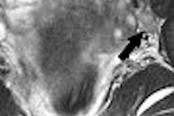
MRI can spot potential complications in Achilles tendon repair and is especially adept at pinpointing cysts or necrosis, according to a presentation at the 2006 RSNA meeting in Chicago.
Dr. Juergen Scheidler and colleagues from the Radiologisches Zentrum Munchen-Pasing in Munich, Germany, conducted their study on 68 patients with partial or full-thickness tears of the Achilles tendon. Of these 68, 47 had surgical repair while the rest were treated nonsurgically.
Scheidler described the three phases of healing after Achilles tendon repair: inflammatory, angiogenesis, and remodeling, all of which are generally reflected on MRI.
For the study, MRI was performed before treatment, then after the onset of therapy at four to six weeks, eight to 10 weeks, and 16 to 18 weeks. The protocol included T2-weighted sagittal FSE fat sequences, axial T1-weighted SE sequences, T2-weighted FSE fat-saturated imaging, and T1-weighted SE fat-saturated imaging with gadolinium.
After four to six weeks, 42 of 68 Achilles tendons demonstrated intermediate to low signal intensity on T1- and T2-weighted sequences, as well as areas of contrast enhancement. This pattern was considered a normal healing process.
At eight to 10 weeks, 41 of 42 continued to heal and displayed decreasing signal intensity, as well as contrast enhancement. At 16 to 18 weeks, 41 patients remained asymptomatic.
One traumatic retear occurred in one patient. In 16 tendons with previous patterns of cyst and/or necroses, the MR patterns did not change and patient symptoms remained. Symptoms persisted and 13 of 16 underwent surgery again, during which necroses were found.
Scheidler concluded that MRI was well-suited to monitor the healing and repair process, and that T2-weighted imaging could serve as an early indicator of outcome. MR results could also show viable repair tissue in cases of hypovascular tendinosis.
An RSNA session attendee asked how long after surgery the abnormally high signal intensity on T2-weighted images would appear. Scheidler said that six weeks after treatment or surgery, the high T2-weighted signal should no longer be present if the tendon was healing properly. However, he acknowledged that a prospective study was needed to take a closer look at MR results from zero to six weeks.
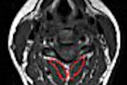
In related research, radiologists at the Karolinska Institute in Stockholm, Sweden, found that MR signal correlated to pain and functional impairment in chronic Achilles tendinopathy.
They found that the severity of pain and functional impairment correlated to increased mean intratendinous signal in the painful tendon on all MR sequences, with significant differences in mean intratendinous signal between symptomatic and contralateral asymptomatic tendons (Acta Radiologica, September 2006, Vol. 47:7, pp. 718-724).
Early detection of potential complication may be particularly important in young athletes, who are more susceptible to rerupturing the Achilles tendon after repair. A study in the American Journal of Sports Medicine found that athletes age 30 and younger were more likely to suffer another rupture, even after successful rehabilitation, especially if they participated in sports with "explosive acceleration" (American Journal of Sports Medicine, January 2005, Vol. 33:1, pp. 119-123).
By Shalmali Pal
AuntMinnie.com staff writer
December 22, 2006
Related Reading
MRI, US take on diagnostic challenge of PAI in soccer players, August 18, 2006
US overcomes x-ray's limits in pediatric ankle fractures, January 23, 2006
Extra pounds add stress to feet and ankles, July 22, 2005
Copyright © 2006 AuntMinnie.com