From the "Greatest Show on Earth" to the some of the greatest hoaxes ever perpetrated, P. T. Barnum knew how to entertain and draw crowds. Even now, more than a century after his death, Barnum's legacy continues to draw interest. In the latest case, a mummy reported to be 2,500 years old takes center stage -- and it's no hoax, according to findings revealed by modern medical imaging.
The 19th century American showman and entertainer Phineas Taylor Barnum (1810-1891) is perhaps best known today for the Barnum & Bailey Circus, and is remembered for his menagerie of sideshow exhibits featuring the Siamese twins Chang and Eng Bunker and the tiny Charles "General Tom Thumb" Stratton. But Barnum was also famous for his great hoaxes like the Fejee Mermaid, which was reported to actually be a fish tail stitched to the body of an ape or monkey.
 |
| "The Barnum & Bailey Greatest Show on Earth: The peerless prodigies of physical phenomena and great presentation of marvelous living human curiosities." A lithographic color print of Barnum & Bailey Circus poster from the Strobridge Lithography Co., Cincinnati and New York, 1899; Library of Congress. |
Now, questions surrounding another piece from Barnum's collection, the Pa-Ib mummy, are beginning to be answered. The mummy's origins, and exactly how it came to be in Barnum's possession, remain a mystery. What is certain is that the mummy was donated to the Barnum Museum in Bridgeport, CT, in 1892 by Barnum's wife after his death, according to the museum. The mummy is purportedly the remains of a priest who served the Egyptian deity Min during the 26th dynasty (circa 664-525 B.C.).
 |
| Pa-Ib in display case at the Barnum Museum in Bridgeport, CT. Photo taken by John Posh, technical director of the Imaging Center at Good Shepherd in Allentown, PA. |
More than 100 years later, imaging and two paleoimaging experts from Quinnipiac University in Hamden, CT, have discovered that the museum's oldest artifact is in fact a real mummy, and not one of Barnum's elaborate hoaxes.
Professors Ronald Beckett, Ph.D., and Gerald (Jerry) Conlogue originally asked the Barnum Museum in 1999 about examining the mummy, but it wasn't until the museum came under new directorship in 2005 that the pair heard back regarding the request.
"The museum contacted us to see if we would be interested in studying their mummy, and since this is what we love to do, we accepted the invitation," Beckett wrote in an e-mail to AuntMinnie.com.
Beckett is a professor of respiratory care and chairperson of cardiopulmonary sciences and diagnostic imaging. Conlogue is an associate professor of diagnostic imaging. They are also the founders and executive directors of Quinnipiac's Bioanthropology Research Institute, and hosts of "The Mummy Road Show," a TV show which aired on the National Geographic Channel that covered 40 episodes in its three-year run and now airs in some international locations in syndication. Between the two, they have investigated more than 500 mummies in countries around the world, and they continue to investigate one to four mummies a year.
In September, Beckett and Conlogue conducted preliminary testing on the Pa-Ib mummy that included endoscopy and light reflectance studies.
"I used an industrial 6-mm Olympus video endoscope, with a halogen light source and a camera control unit. Images are captured on digital videotape (Canon Elura) and introduced into iMovie (Apple) on a MacBook Pro laptop, where I can capture a frame or a movie," Beckett wrote. Light reflectance is a new technique that uses a reflectance probe (Ocean Optics) and instrumentation, he explained. Reflectance signatures for various tissue types were captured and compared.
Earlier this month, the mummy was transported to Advanced Radiology Consultants in Fairfield, CT, for further imaging with computed tomography. With help from the practice's imaging specialists, a whole-body CT scan was performed with a 32-slice multidetector-row CT scanner (Aquilion 32, Toshiba America Medical Systems, Tustin, CA).
 |
| Above: Quinnipiac University professors Jerry Conlogue and Ronald Beckett along with Kathy Maher, executive director and curator for the Barnum Museum, examine Pa-Ib prior to transport on October 11 to Advanced Radiology Consultants in Fairfield, CT. Below: At the Barnum Museum, the mummy is moved for wrapping and transport. Bottom: Beckett and Conlogue have Pa-Ib delicately wrapped in special paper for protection and preservation and prepared for transport to the Advanced Radiology Consultants facility for CT and MRI scans. Photos taken by John Posh, technical director of the Imaging Center at Good Shepherd in Allentown, PA. |
 |
 |
 |
| The mummy was respectfully transported in a coffin to the imaging facility by Michael Parente and Jack Migliore from Parente-Lauro Funeral Home of Bridgeport, CT. Photo taken by John Posh, technical director of the Imaging Center at Good Shepherd in Allentown, PA. |
One of the most surprising findings on the CT data is that the mummy appears to be female due to its pelvic structure and signs of arthritis in the area, a possible indication of having given birth, according to experts at Advanced Radiology. Despite the findings contradicting the long-held belief that the mummy was male, the remains could still be those of an Egyptian priest as claimed, as women also held that position, according to Barnum Museum's executive director and curator Kathy Maher.
 |
| From left to right: Dr. Ruben Kier, chairman of the board for Advanced Radiology Consultants; Beckett; Amy Kovak, senior CT technologist at Advanced Radiology; and Maher prepare to enter the mummy into the Toshiba Aquilion 32 CT scanner. Image courtesy of Michelle Cotton, Advanced Radiology Consultants. |
The mummy also contained three to five "organ packets ... within the thoracic and abdominal regions" and "worn teeth from sand in the food," as well as a "huge opening into the cranial vault made to scramble the brains for removal" during the mummification process, according to Beckett. The mummy may even be from an older dynasty than was previously thought.
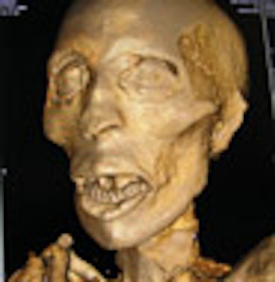
 |
| Above and below: 3D reconstructions from CT data of the mummy's head, scanned at 100 kVp, 100 mA, 750 msec rotation speed, 75 mAs, and a slice thickness of 0.5 mm. Images reconstructed using Vitrea 2 advanced visualization software (Vital Images, Minneapolis, MN). Images courtesy of Michelle Cotton, Advanced Radiology Consultants. |
 |
 |
| Above: The thoracic and pelvic regions of the mummy. Below: A lateral view of the regions. Bottom: The pelvis and legs. Images courtesy of Michelle Cotton, Advanced Radiology Consultants. |
 |
 |
The mummy also underwent an MRI scan, but the imaging resulted in no additional findings, of course, due to the artifact's dry condition.
The CT images need further review, but the preliminary findings have confirmed the authenticity of the Barnum Museum's Pa-Ib mummy, ending the speculation that it's just another hoax by one of the 19th century's greatest showmen.
Beckett and Conlogue are working on writing up their research, which will be reported at the VI World Congress on Mummy Studies, to be held February 20-24, 2007, on the Canary Islands of Spain, according to Beckett.
By Heather Hokenson
AuntMinnie.com staff writer
October 31, 2006
Related Reading
Scan artist: Radiologist uses CT to reveal mystery of antiquities, October 25, 2005
CT helps unwrap mummy mystery, March 29, 2005
MDCT solves 400-year-old mystery, November 29, 2004
Mummy dearest: CT helps archaeologists dig deeper, April 23, 2003
Mummyography: Images reveal secrets of ancient remains, December 20, 1999
Copyright © 2006 AuntMinnie.com