A new study by Italian researchers has found that digital radiography (DR) delivers lower radiation dose to patients than either conventional x-ray or computed radiography (CR) systems. In comparison to CR, DR delivers nearly half the radiation dose of the competing digital modality.
The researchers said they began to study the radiation dose characteristics of the three different radiography modalities after installing a new DR system in the accident and emergency department at their facility, Orsola-Malpighi Hospital in Bologna. The department conducts about 50,000 standard radiography exams annually out of a total of 400,000 performed overall at the hospital. The study was published in the November issue of the British Journal of Radiology (November 2006, Vol. 79, No. 947, pp. 899-904).
In discussing their decision to convert to digital x-ray, the researchers cited advantages such as its greater dynamic range, wider exposure latitude, and image postprocessing capabilities, as well as lower film costs and the ability to distribute images to clinicians easily. The researchers said they felt DR in particular was well-suited to the trauma environment due to its ability to conduct procedures of critically ill patients relatively easily.
The department in 2003 had replaced one of its three conventional radiography rooms with CR readers (DirectView CR-850 and CR-900, Eastman Kodak Health Group, Rochester, NY), and in 2004 replaced another room with a DR unit (Axiom Aristos FX, Siemens Medical Solutions, Erlangen, Germany). The remaining conventional film-screen radiography system used Kodak T-Mat G/RA film and a Trimax regular screen (nominal speed class 400).
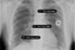
The researchers chose to compare the radiation dose received by patients undergoing six standard radiography exams using the three modalities in 10 projections, including anteroposterior (AP) abdomen, posteroanterior (PA) chest, lateral (LAT) chest, AP lumbar spine, LAT lumbar spine, LAT lumbosacral joint, AP pelvis, AP skull, LAT skull, and AP urinary tract. The group measured radiation dose using the entrance surface dose (ESD) and effective (E) dose techniques.
Although manufacturers of digital x-ray systems generally recommend the use of low x-ray tube potentials, the researchers said that with their digital systems they used higher tube potentials and lower mAs to reduce patient radiation dose. Digital postprocessing tools can help compensate for the lower contrast produced by images created with higher tube potentials, the researchers stated.
The group first calculated entrance surface dose and effective dose by measuring x-ray tube output for every tube voltage, then applied guidelines from the European Commission and the U.K. National Radiological Protection Board (now the Radiation Protection Division of the Health Protection Agency) for deriving ESD and E.
Both entrance surface dose and effective dose varied greatly depending on the type of procedure being conducted and the modality used. DR had the best performance in LAT lumbosacral joint exams, with an ESD that was 66% lower than film-screen radiography and 68% lower than CR. DR's ESD radiation dose was higher than film-screen radiography only in one category, AP skull exams, with DR producing 10% more radiation than film-screen radiography for that application and just 2% less than CR for the same exams.
For radiation dose as measured by effective dose, DR performed best in pelvic AP exams, with an E that measured 43% below film-screen radiography and 48% below CR. DR's biggest advantage over CR was in lumbar spine AP and lateral exams, where it had 62% lower radiation dose.
For complete examinations, DR's effective dose was about 29% lower than film-screen radiography and 43% lower than CR, according to the researchers.
The authors reported that their findings on radiation dose were about in line with previous studies. Their findings led them to state that "doses with the use of CR systems are approximately the same as those for a 300-speed (film-screen) system." Image optimization protocols are important, however, as there are big differences in image quality for similar-speed systems, they noted.
Also, CR's higher radiation dose for some exams was mitigated by its greater dynamic range, which meant that fewer exams had to be repeated. The authors also cautioned that CR's higher dose characteristics should be put in perspective. A facility would have to conduct complete chest exams on nearly 1 million patients to produce one additional health defect (such as cancer) compared to film-screen radiography, and two additional health defects compared to DR, based on International Commission on Radiological Protection (ICRP) data on cancer risk.
The authors concluded by citing additional advantages of DR systems, which include shorter image acquisition times compared to CR and film-screen radiography, increased patient throughput, and the rapid availability of images. Users may be able to reduce DR radiation dose further thanks to the technical characteristics of the technology.
"The image quality characteristics of flat-panel technology are theoretically better than those of (film-screen radiography) and CR because at a comparable resolution the (DR) has a higher detective quantum efficiency (DQE)," they wrote. "This advantage can be utilized in reducing the patient dose whilst maintaining image quality and without changing the signal-to-noise ratio. The appropriateness of this approach would, however, require verification."
By Brian Casey
AuntMinnie.com staff writers
November 7, 2006
Related Reading
U.S. hospitals find ways to take the digital x-ray plunge, October 12, 2006
Digital radiography slowly, but surely, makes its mark, July 25, 2006
Digital x-ray unreliable for evaluating bone healing, May 31, 2006
Asian digital x-ray market primed for growth, February 17, 2006
Copyright © 2006 AuntMinnie.com