One of the first virtual colonoscopy studies to use a 64-slice CT scanner with computer-aided detection (CAD) concluded that the software found lesions radiologists missed, missed lesions radiologists found, and was helpful overall.
The challenges of virtual colonoscopy practice include results that are highly reader-dependent, a steep learning curve, and lengthy interpretation times, said Dr. Anno Graser from the Grosshadern Campus of the University of Munich, Germany, at the 2006 European Congress of Radiology (ECR).
"The benefits of CAD include a potential increase in new readers' diagnostic performance and a decrease in the variability of diagnostic performance among human readers -- especially those who are less experienced," Graser said. "And CAD can decrease human readers' perceptual errors."
The study by Graser, Dr. Christoph Becker, Dr. Maximilian Reiser, and colleagues tested the performance of a virtual colonoscopy CAD system (syngo Colonography CT with Polyp-Enhanced View [PEV], Siemens Medical Solutions, Erlangen, Germany) that was investigational at the time of the study but has since been approved by the U.S. Food and Drug Administration for clinical use, Graser said.
 |
| The syngo Colonography CT CAD interface. All images courtesy of Dr. Anno Graser. |
A cathartic bowel prep using polyethylene glycol plus four 45-mg bisacodyl tablets was administered 24 hours before scanning. The subjects also underwent same-day conventional colonoscopy, which served as the reference standard.
In all 105 subjects, including 42 with polyps and 63 negatives (52 men, 53 women; mean 59 years) underwent multidetector-row CT (MDCT) colonography, or virtual colonoscopy, for colon cancer screening on a 64-slice scanner (Somatom Sensation 64, Siemens Medical Solutions).
The subjects were scanned in both prone and supine positions. The investigators did not use fluid or fecal tagging, which had not yet been evaluated with the system. Data were acquired at 0.6-mm collimation, 120 kVp, 40 (reference) mAs, and dose modulation in the x, y, and z axes. The slice thickness was 0.75 mm, with 0.5-mm overlapping reconstructions, Graser said.
Axial slices 1-mm thick were reconstructed for 3D visualization. Two expert readers interpreted the data using a primary 3D endoluminal approach. The CAD system was used as a second reader to assess its sensitivity for detecting polyps, as well as the number of false-positive findings per dataset.
The reconstructed images were read on a 3D Siemens syngo Colon postprocessing workstation by an expert reader with experience in reading more than 700 virtual colonoscopy cases.
In all, 98 polyps were found in 42 patients, including 51 small (< 5 mm) lesions, 32 medium (6-9 mm) lesions, and 15 large (> 9 mm) lesions. CAD produced an average 2.2 false positives per dataset.
The mean running time of the CAD software was 4.0 minutes per dataset, which consisted of approximately 1,700-2,000 images per patient including prone and supine data, Graser said.
CAD's sensitivity was 51% for small polyps, 94% for medium, and 87% for large (70% overall), lower than the radiologists' rates of 84% for small, 94% for medium, and 93% for large (80% overall).
 |
| CAD performed best in the detection of medium-sized lesions (6-9 mm), and worst for lesions 5 mm and smaller. |
"CAD was significantly worse in detection of small lesions," Graser said. "But it was pretty high in the medium-sized lesions and in the large lesions."
 |
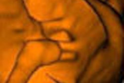
| CAD and radiologist's marks concur for detection of a medium-sized polyp. |
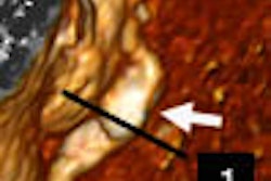
Still, CAD missed two large polyps, including a malignancy. Pointing to the CT image of the mass, Graser noted its "large ridge-like structures, more like folds than caplike bulbous polyps. Maybe that was the reason CAD missed the lesion, which was easily detected by the radiologists. It was a transmural lesion. You see the lymph nodes up here -- definitely something you would not miss." CAD also missed a large adenoma that was harder to see.
 |
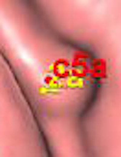
| Above, CAD missed a large rectal mass. Below, CAD missed a 3-cm flat tubulovillous adenoma that was also missed by the radiologist. |
 |
"It was a flat tubular adenoma that I actually missed and so did CAD," Graser said. "It looks like a fold in the endoluminal view, and you see the fold in 2D as well. Its texture is different from this adjacent fold, and this was why the endoscopist did detect it and it was resected."
Despite the misses, CAD may add incremental value even to the expert reader, since in many cases the algorithm detects different patterns than a human reader would find, Graser said. For the CAD software, "it doesn't really matter if a lesion is located between two folds or sitting on a fold," he said.
CAD is likely to improve in its ability to detect large masses that can elude detection now; however, flat adenomas will continue to be problematic, Graser predicted.
By Eric Barnes
AuntMinnie.com staff writer
May 12, 2006
Related Reading
VC CAD helps junior readers catch up, May 4, 2006
VC CAD improves results for readers at all levels, April 7, 2006
Colon CAD helps expert readers, March 16, 2006
VC CAD found equivalent to colonoscopy in screening population, December 21, 2005
New VC reading schemes could solve old problems, October 13, 2005
Copyright © 2006 AuntMinnie.com