Three-dimensional sonography of the endometrium and adjacent myometrium can yield additional useful information over traditional 2D studies in almost a third of routinely performed examinations, according to a study from Vanderbilt University Medical Center in Nashville, TN.
"The coronal image of the endometrium and adjacent myometrium is an easily obtainable adjunct to any conventional transvaginal pelvic sonogram," said lead author Dr. Rochelle Andreotti. "Additional useful information may be obtained in three out of 10 total exams, and five out of 10 exams that are abnormal by 2D imaging."
Andreotti presented the Vanderbilt research during a scientific session at the annual convention of the American Institute of Ultrasound in Medicine (AIUM) in Washington, DC, in March.
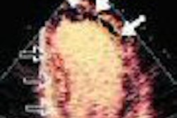
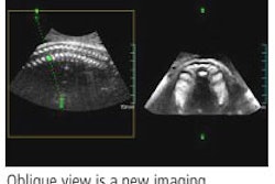
Traditional 2D sonography views of the uterus and endometrium are usually limited to sagittal and transverse views of the uterus. But with volume imaging, the uterus and endometrium can now be imaged in the coronal plane, Andreotti said.
Investigators have demonstrated additional findings on coronal images that can't be appreciated on traditional views, the most obvious being uterine anomalies, she said. The relationship of abnormalities to the endometrial cavity has also been under scrutiny.
The Vanderbilt researchers attempted to characterize the types of additional information that can be obtained in the coronal plane and with what frequency, evaluating a series of patients presenting for pelvic sonography with routine 2D as well as 3D reconstructed images of the endometrium in the coronal plane.
Ninety randomly selected patients presenting for transvaginal pelvic sonography were imaged by experienced sonographers according to standard 2D protocol using either a 3D 9-3 MHz broadband transvaginal transducer or an 8-4 MHz broadband transvaginal transducer.
Following the 2D exam, a 3D uterine volume was acquired using either an automated or manual sweep in the sagittal plane and reconstructed in the coronal plane, according to Andreotti. Multiple images were obtained through the anterior-posterior thickness of the endometrium.
Images were reviewed retrospectively by experienced sonologists. The endometrium and surrounding myometrium were evaluated for architecture, masses, relationship of masses to the endometrial cavity, and the anatomic configuration of the cavity, she said.
Ninety-one studies were obtained in the 90 patients. Twenty-eight (30.8%) of the studies demonstrated additional findings in the coronal plane, and two additional findings were seen in three studies. No additional findings were obtained in 63 (69.2%) cases.
The additional findings included uterine anomalies in eight cases, better delineation of endometrium in seven, better delineation and location of polyps in six, better location of leiomyomas in five, cystic areas in myometrium in three, and confirmation of intrauterine device (IUD) location in two.
The researchers then divided the findings into two categories: patients with a normal uterus and endometrium on 2D sonography and those with abnormal findings.
On 2D sonography, 42 of 91 had normal findings. In this group, additional findings using 3D reconstruction in the coronal plane were gained in only two (5%) patients, one with confirmation of IUD location and one with an arcuate uterus.
Of the 49 cases with abnormal findings on 2D transvaginal sonography, additional findings from 3D reconstruction of the coronal plane were found in 26 (53%) of cases.
"Additional useful information will not likely be obtained if the 2D exam of the uterus is normal," she said.
By Erik L. Ridley
AuntMinnie.com staff writer
May 5, 2006
Related Reading
3D ultrasound predicts fetal pulmonary hypoplasia, April 21, 2006
Ultrasound alters management of infants with genitourinary disorders, October 14, 2004
Embryo and Fetal Pathology: Color Atlas with Ultrasound Correlation, October 13, 2004
Three-dimensional ultrasound shows utility in fetal lung volume measurements, March 18, 2004
Part II: The psychological impact of 3-D ultrasound on pregnant women, December 30, 2003
Copyright © 2006 AuntMinnie.com