The increasing utilization of cross-sectional modalities and digital mammography will drive breast imaging practices to switch to soft-copy reading within the next five years, according to Dr. Mitchell Schnall, associate chair for research at the Hospital of the University of Pennsylvania in Philadelphia. And as part of this transition, mammographers will find 3D visualization tools to be a growing part of their daily activities.
"Our current concept of breast cancer imaging and screening is going to get much more complicated due to the implementation of other modalities, including some that are three-dimensional, and even the evolution of traditional mammography into 3D through tomosynthesis," he said. "Those that practice in breast imaging need to learn about more sophisticated imaging modalities, workstations, and computers, because they are going to need those."
Schnall spoke about the application of 3D visualization technology for the display of breast images during a session at the 2005 RSNA meeting in Chicago and during an interview with AuntMinnie.com.
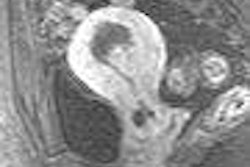
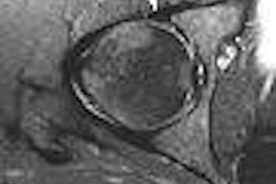
Schnall believes that 3D visualization tools used for the display of 3D breast images represent new learning and significant adaptation challenges for radiologists, even with the commercial software currently available. Presently, MRI is the only 3D breast imaging that is routinely performed in 3D. (This type of 3D image represents the construction of multiple 2D images into an image in three dimensions, and is to be differentiated from volumetric 3D rendering.)
In spite of the steadily increasing use of breast MRI, navigating the complex set of data generated by a breast MRI scan is an obstacle to its integration into a typical breast imaging practice. More and better software is needed to make data interpretation easier and to facilitate the referencing of findings among MRI, ultrasound, and mammography exams, Schnall said.
The value of 3D breast images is their ability to better aid the detection of invasive breast cancer than mammography, particularly with respect to the imaging of dense breasts. Numerous clinical trials conducted over the past two decades have shown that contrast-enhanced MRI breast images detect early-stage breast cancers such as ductal carcinoma in situ (DCIS) and small node-negative cancers with a sensitivity level of 50%.
Breast MRI is also useful in determining the extent of cancer. The technology provides a noninvasive method for assessing the response of a tumor to chemotherapy, and is used for postinterventional therapy monitoring and to detect any local recurrence of cancer.
Breast MRI acquires a 4D dataset consisting of three spatial dimensions and one time dimension. It's difficult to manage and visualize 4D datasets, however, even though available FDA-cleared visualization software automates many tasks, Schnall said. Because of this, the most prevalent 3D visualization technique used by radiologists is rapid scrolling through the 3D dataset in a stack mode as a series of 2D images, he said.
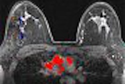
"The problem of analyzing the 4D datasets is to reduce this to a scenario that can be solved," Schnall said. "Time dimension can be obtained by either pointing to locations on the 2D images that provide a plot of what happened over time to create a time-intensity curve, or color coding with some overlay can be used that gives a perimeter extracted from the curves. This provides such information as how rapidly areas take up contrast and how rapidly contrast washes out, and from this make a diagnostic interpretation."
Image subtraction to detect enhancement is automated by software, creating subtraction image sets created from acquisitions over the first two minutes. Registration of postcontrast images to the precontrast image set to reduce subtraction artifacts is another automated function, he said.
"The most challenging part of visualization and display of a 3D MRI exam is the integration of kinetic data with anatomic images," Schnall said. "Clinicians have been very resourceful, and there are many accepted approaches, including qualitative assessment of the time intensity curve."
There is no conclusive evidence that quantitative assessment and modeling of the kinetic curve, which is difficult to generalize, performs better than the qualitative assessment, Schnall said. Color overlays represent an appealing way to combine the time course and anatomic data.
Commercial workstations today are equipped with support tools that classify the time-course curve as plateau, washout, or persistence on a pixel by pixel basis. These tools provide the radiologist with a rapid way to scan the dataset and identify areas of concern based on time-course data.
Value of 3D reconstruction
While 3D volumetric reconstruction of the breast is an exciting visual concept, it offers little diagnostic value, and is of little value to breast surgeons, Schnall said. Because all the diagnostic information is contained in the sequential 2D slices from which a 3D volume image is made, a 3D reconstruction will contain no additional information, and in the process of its creation may show less. Its benefit is to demonstrate an interpretation to clinicians and patients.
The lack of anatomic landmarks of the breast limits the usefulness of 3D volume renderings for breast surgeons.
"Because the breast can be positioned in many different ways, it is difficult to translate the information on the image to the breast so that a surgeon can remove only the tissue that is necessary," Schnall said.
Software innovations that can align 3D breast MRI images to the operative field and to more accurately position noninvasive treatments that kill a tumor without surgery are needed and will undoubtedly be forthcoming in this decade, he said.
Future 3D technologies
Also on the horizon is full-field breast tomosynthesis, which acquires images of a stationary compressed breast at multiple angles. These images are then reconstructed into a series of individual 3D slices. Like CT, these slices are displayed as individual images in a cine loop.
Allowing for reducing or eliminating tissue overlap, tomosynthesis can co-register its images with a digital mammogram using the same compression. Objects become correlated in the two image sets, and the location of a lesion in a tomosynthesis slice can determine its true coordinates in a breast.
The learning curve for radiologists to interpret images generated from tomosynthesis will be easier because they have the same characteristics as digital mammography, Schnall said.
3D breast ultrasound is being developed in research settings. Techniques for registration of 3D datasets are permitting more accurate measurement of tumor volume and comparison of changes in the size of masses over time, he said.
PET and scintimammography also produce 3D images. PET is employed today for restaging and evaluating recurrent breast cancer, while scintimammography is being used in conjunction with mammography for detecting breast cancer in patients with dense breasts, with breasts that have palpable lesions, and with architectural distortions from prior biopsy, surgery, or radiation therapy, Schnall said.
These technologies will be limited in their use for breast imaging, however, due to their diagnostic limitations and their expense, he said.
What do advances in 3D breast imaging portend for the breast imaging center? Radiologists who currently limit their interpretations to mammography and wish to offer cutting-edge technology will need to become proficient in 3D breast MRI interpretation, Schnall said.
The technology may lead to greater specialization as well as remote interpretation by expert 3D mammographers at centralized facilities. In either case, 3D breast imaging will undoubtedly lead to significant changes in the process of diagnosing breast cancers, Schnall said
By Cynthia Keen
AuntMinnie.com contributing writer
April 11, 2006
Related Reading
Swedish start-up XCounter pursues 3D mammography, March 20, 2006
Technological advances drive new 3D CPT codes, February 21, 2006
3D boosts efficiency of transvaginal sonography, February 8, 2006
Imaging poised to transform future of medicine, February 2, 2006
High resolution, fast acquisition give 3D MRA edge in abdomen, January 20, 2006
Copyright © 2006 AuntMinnie.com