Contrast-enhanced ultrasound (CEUS) can help diagnose indeterminate small hepatic lesions, according to researchers from the University of California, San Diego (UCSD).
While CT and MRI are good tools for diagnosing liver tumors, the modalities can't characterize a subset of lesions, especially small ones, according to Dr. Yuko Kono, an assistant clinical professor at UCSD. CEUS, however, has been shown to be accurate in characterizing liver lesions.
To assess the accuracy and role of CEUS in diagnosing small lesions that were indeterminate on CT and/or MRI, the UCSD researchers studied 55 patients with indeterminate early enhancing lesions smaller than 3 cm in diameter that were detected by contrast-enhanced CT and/or contrast-enhanced MRI. Kono presented the research during a scientific session at the annual meeting of the American Institute of Ultrasound in Medicine (AIUM), held last week in Washington, DC.
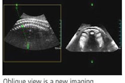
Depending on availability, the UCSD team employed Imagent (Imcor Pharmaceuticals, San Diego), Optison (GE Healthcare, Chalfont St. Giles, U.K.), or Definity (Bristol-Myers Squibb Medical Imaging, North Billerica, MA) ultrasound contrast agents with an intravenous multiple bolus injection. Scanning was performed using a Sonoline Elegra or Antares ultrasound system (Siemens Medical Solutions, Malvern, PA), with phased-inversion harmonic imaging (PIHI), contrast pulse sequencing (CPS), real-time and intermittent imaging, and a 3.5-MHz curved transducer.
The researchers used CEUS to evaluate tumor vessels, as well as the pattern and degree of enhancement on arterial, portal, and equilibrium phases on real-time low MI, intermittent, and long-delay imaging, Kono said.
A final diagnosis was available in 35 patients, 13 via histology and 22 by imaging follow-up (average of 26 months). Of these, 17 patients had one lesion, 11 patients had two lesions, and seven patients had more than two lesions.
Prior to receiving CEUS, 24 patients received triple-phase CT studies, 26 underwent dynamic MRI exams, and 15 received both imaging studies. Twenty-three patients were diagnosed as cirrhotic, and 12 had no liver disease, Kono said.
Of the 23 cirrhotic patients, 14 had benign lesions and nine had malignant lesions, all of which were hepatocellular carcinoma (HCC). The 12 noncirrhotic patients had 11 benign lesions and one malignant lesion, a breast cancer metastasis.
Noncontrast ultrasound produced a sensitivity of 78% (five false negatives) and a specificity of 92% (one false positive) for detecting lesions, Kono said. CEUS, however, yielded a 91% sensitivity (two false negatives) and 100% specificity for lesion detection.
For the diagnosis of malignancy, CEUS turned in 100% sensitivity (10 of 10 malignant lesions) and 92% specificity (25 of 27 benign lesions), Kono said.
"CEUS is helpful in diagnosing indeterminate small hepatic lesions in both cirrhotic patients and patients without liver disease," she said.
By Erik L. Ridley
AuntMinnie.com staff writer
April 3, 2006
Related Reading
Detection of liver metastases with contrast-enhanced ultrasound, March 23, 2006
Contrast-enhanced US useful for differentiating focal liver lesions, January 19, 2006
Contrast-enhanced ultrasound aids in grading liver disease, December 1, 2005
Ultrasound contrast enhances liver imaging, May 11, 2005
Exam technique critical in liver ultrasound, October, 22, 2004
Copyright © 2006 AuntMinnie.com