Thin-section multidetector-row CT, which does such a fine job of visualizing calcified atherosclerotic plaque, is all but blind when it comes to differentiating two important but morphologically distinct types of noncalcified coronary plaque.
The limitation is important considering that fibrous noncalcified plaques are generally considered stable and therefore less dangerous, while soft plaques with a large lipid core are far more vulnerable to rupture. Knowing the predominant soft-plaque type can therefore be important to patient management.
"The basic question is: Can we characterize plaque morphology based on the concept of vulnerable plaque being characterized by a large lipid core?" asked Dr. Friedrich Knollman in a presentation at the 2005 RSNA meeting in Chicago.
In search of an answer, "we set out to quantitatively assess plaque components and look for a lipid core based on CT attenuation," he said. "And to do that we chose a postmortem investigation in the belief that histology is the appropriate standard for such an investigation."
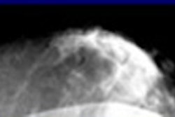
Knollman and colleagues at the Georg-August University of Göttingen in Germany prospectively evaluated 30 human hearts that had been excised from cadavers. Prior to imaging, the coronary arteries were filled with a barium sulfate-based contrast medium and depicted with a 16-row MDCT scanner (LightSpeed Pro 16, GE Healthcare, Chalfont St. Giles, U.K.). Data were acquired at 120 kVp. The slice thickness was 0.6 mm, and overlapping reconstruction, depending on the orientation of the vessel in z-axis, yielded a voxel size of 0.4 mm to 0.6 mm, he said.
"In this postmortem setting we didn't have any motion -- we had hearts that were not beating," he said. "So we eliminated motion artifacts and had an ideal setup to look at the morphology, acknowledging that in the clinical setting you would also have to deal with motion."
The images were reformatted perpendicular to the axis of the three main coronary arteries (LAD, RCx, RCA; Advantage Workstation 4.2 software, GE Healthcare), and analyzed by determining the attenuation profile of the resultant coronary cross sections (ImageJ software, National Institutes of Health, Bethesda, MD). Then the attenuation measurements of plaque components were compared with light microscopy of anatomically matched pathologic specimens.
According to the results, epicardial fat showed attenuation between -120 and 23 HU (median, -71 HU), while the attenuation of the contrast-filled lumen ranged from 93-625 HU (median, 310 HU). Calcified atherosclerotic plaque could be easily identified by its much higher attenuation of 333-1,944 HU (median, 1,089 HU).
"Not surprisingly, calcification could be identified very well with high accuracy, and could be differentiated from the luminal contrast agent also, which in turn could be differentiated very well from the noncalcified plaque component," Knollman said.
On the positive side, the difference in attenuation between the two types of soft plaques was statistically significant (ANOVA, p < 0.0001).
The uncalcified atherosclerotic plaque had an attenuation of 26-168 HU (median, 53 HU). However, a subclassification of uncalcified plaque components as either lipid rich (27-67 HU; median, 48 HU) or fibrous (37-133 HU; median, 67 HU) showed substantial overlap between the two, and therefore did not allow for consistent differentiation.
"We can identify calcified and noncalcified coronary atherosclerotic plaques; however, the detection of the necrotic lipid core, which is soon to be the hallmark of a vulnerable lipid, remains uncertain," Knollman said. The ROC curve for differentiating fibrous from lipid plaque showed that "sensitivity and specificity in an ideal setting won't be much better than 80% sensitivity and less with (slightly) higher specificity," he said.
On thin-section 16-slice MDCT, the further differentiation of uncalcified plaque components is not feasible, and the identification of plaque vulnerability based on the size of its lipid core remains uncertain even when motion artifacts are eliminated, he concluded.
Yet the study techniques are potentially useful. The identification of thresholds using profile analysis can be used to perform plaque volumetry, providing quantitative information on total plaque volume, total calcified and noncalcified plaque volume, and the ratio of calcified to noncalcified plaque.
Further scanning was performed by varying the tube voltage, and the analysis will be available at a later date, Knollman said.
By Eric Barnes
AuntMinnie.com staff writer
January 10, 2006
Related Reading
Traditional risk factors may fail to identify women with atherosclerosis, December 19, 2005
Long-term anticoagulant users show elevated coronary, aortic calcium, December 2, 2005
Caution: U.S.A. may be hazardous to your health, October 3, 2005
Coronary calcium screening seen useful beginning between age 40 and 50, September 23, 2005
Low-dose CT calcium scores near-equivalent of higher dose, September 20, 2005
Copyright © 2006 AuntMinnie.com