Integrating real-time 3D into a 2D PACS viewing environment can solve a host of interpretation challenges and yield significant workflow gains, according to a presentation at the Society for Computer Applications in Radiology (SCAR) meeting in Orlando, FL.
"It is becoming increasingly useful to have an integrated workstation that seamlessly integrates the conventional 2D viewing with advanced 3D postprocessing capability," said Dr. Vamsi Narra, an assistant professor of radiology at the Mallinckrodt Institute of Radiology (MIR) in St. Louis.
Before pursuing an integrated 2D/3D viewing environment for its body MRI service, MIR used its PACS network (Sienet and MagicView, Siemens Medical Solutions, Malvern, PA) primarily for 2D image viewing, with 3D capability available largely through a dedicated 3D workstation (Vitrea, Vital Images, Plymouth, MN).
The institution performs an average of about 30 body MRI and MRA cases a day, and wanted to bring the power of real-time 3D to problem solving in daily interpretations. By doing so, MIR hoped to streamline workflow and gain access to postprocessed images tailored to specific clinical questions, Narra said. MIR also wanted to be able to seamlessly share the information with clinicians.
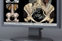
The revamped workflow begins with the facility's MR scanners sending datasets to both the PACS and an integrated workstation containing integrated 2D and 3D capability. Radiologists use an application (SoftRead, Vital Images) to view the MR studies in 2D mode, and at any point, can bring up the current series in 3D mode with a single click, Narra said.
As a result, radiologists are able to immediately employ 3D tools such as maximal intensity projection, minimal intensity projection, multiplanar reconstruction, volume rendering, and surface shaded display, he said.
Radiologists can also take "snapshots" of any clinically interesting image, which can be posted on the PACS system, an intranet viewing system for clinicians, and printed or e-mailed to a referring clinician. Annotations can be made as well.
"3D can really impact clinical real-time interpretation of these cases," Narra said
Success
Implementation of the new workflow model began 18 months ago, initially with one radiologist using the system. MIR's 13 radiologists had the option of retaining the old workflow model of using the dedicated 3D workstation or converting to the new integrated 2D plus 3D approach.
Within eight months, 90% of the radiologists had begun using 2D/3D readout, a figure that reached 100% after one year. Pressure from residents and clinician preferences forced reluctant radiologists to finally change, Narra said.
"Clinicians, when they come in and review cases, they would rather have them to review in 3D," he said.
Initially, 3D processing was first applied at MIR to formal 3D reconstruction cases such as planning for endoluminal aneurysm grafts. These cases require a specified set of 3D measurements, and these studies are still processed by a technologist to obtain reconstructions and measurements for the radiologist to review, Narra said.
However, the ready availability of 3D has opened the door for an increased role in a wide variety of routine clinical cases, significantly improving understanding of pathology via real-time 3D visualization of vascular structures and soft tissue structures, he said.
By Erik L. Ridley
AuntMinnie.com staff writer
June 17, 2005
Related Reading
Complex cardiothoracic surgery benefits from 3D imaging, June 13, 2005
Real-time 3D/MPR image processing feasible over Internet, June 3, 2005
3D diagnosis and planning bring orthodontists closer to 'anatomic truth,' June 2, 2005
3D: Rendering a new era, May 2, 2005
4D ultrasound shows promise for improving fetal echocardiography, April 6, 2005
Copyright © 2005 AuntMinnie.com