It may seem like little more than a technicality, but proper image segmentation on computer-aided detection systems (CAD) can directly influence the success of this technology. When done correctly, segmentation excludes background regions from analysis, improving accuracy and reducing computation time.
But improper segmentation can result in missed information, warned researchers from Duke University in Durham, NC. In the case of breast imaging, a malignant lesion in a breast region that was excluded by the segmentation process will not be identified by the CAD system, they explained. The group, led by Dr. Jay Baker, tested the accuracy of CAD segmentation in the screening mammograms of 507 women over a two-month period.
"The specific results of our study both illustrate the effect of the segmentation technique on CAD analysis and demonstrate the importance of attention to segmentation accuracy during clinical implementation of CAD," they wrote (Radiology, March 15, 2005).
For this study 2,020 standard film-screen mammographic views (Mammomat 3000, Siemens Medical Solutions, Malvern, PA; MinR-2000, Eastman Kodak, Rochester, NY) were included and digitized at 50-µm resolution and 12-bit grayscale. CAD analysis was then performed (ImageChecker M1000, version 3.2, R2 Technology, Sunnyvale, CA).
Baker and co-author Dr. Eric Rosen reviewed each case for overall density of the breast parenchyma, and assessed the accuracy of CAD segmentation. Near-perfect segmentation indicated that skin contour and all breast parenchyma were identified. Acceptable segmentation meant that all of the parenchyma was included, but not all of the nonparenchymal adipose tissue was included. Segmentation was deemed unacceptable if more than 5% of actual parenchyma was excluded.
 |
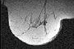
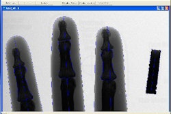
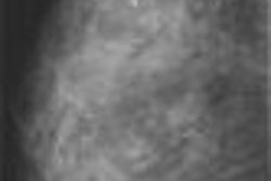
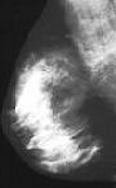
| Bilateral CAD images in a 54-year-old woman demonstrate unacceptable segmentation of breast parenchyma. Left: Craniocaudal CAD image. CAD segmentation excluded 50% of breast parenchyma (arrows). Right: Mediolateral oblique CAD image demonstrates near-perfect segmentation. Baker JA, Rosen EL, Crockett MM, Lo JY, "Accuracy of Segmentation of a Commercial Computer-aided Detection System for Mammography," Radiology 2005:235(2):385-390. doi:10.1148/radiol.2352040899. Published March 15, 2005; accessed April 7, 2005. |
Overall, segmentation was near-perfect or acceptable in 96.8% of the cases and unacceptable in 6.2%. Similar results were achieved for mammograms that were categorized as fatty replaced or scattered fibroglandular tissue (98.2% near-perfect or acceptable for the latter). The area of excluded tissue averaged 24.7%.
In heterogeneously dense breasts, only 92% of the exams were considered near-perfect or acceptable; 8% were unacceptable. "This level of unacceptable segmentation for heterogeneously dense breasts is over five times the 1.4% unacceptable rate for the other three categories of breast density combined," the authors stated.
"Our findings ... alert practicing radiologists to the issue of segmentation, and demonstrate that dependence of segmentation on breast tissue density," they added.
Baker's group acknowledged the limitations of their study -- they did not directly address the issue of faulty segmentation with regard to causes, reproducibility, sensitivity, and specificity. Nevertheless, their research highlighted that an inaccurate segmentation process can result in the exclusion of a substantial portion of breast tissue.
By Shalmali Pal
AuntMinnie.com staff writer
April 26, 2005
Related Reading
CAD offers promising second opinion, even in dense breasts, March 15, 2005
CAD systems show double-digit growth in Europe, November 23, 2004
CAD may unmask unsuspected cancers in diagnostic mammo, October 27, 2004
Copyright © 2005 AuntMinnie.com