Differentiating high-grade gliomas from gray-matter structures with F-18 FDG-PET has been problematic for clinicians because of the brain's avidity for the radiotracer. A recent study from the University of Washington School of Medicine in Seattle presents evidence that extending the interval between FDG administration and PET data acquisition improves the delineation of gliomas from gray matter.
"Several studies have explored the usefulness of delayed or dual-time-point F-18 FDG-PET in improving detection of brain metastases and tumors outside the brain, distinguishing malignant from benign lesions, distinguishing malignant from inflammatory lesions, or defining the time for optimal imaging of tumors," the authors wrote in the Journal of Nuclear Medicine (October 2004, Vol. 45:10, pp. 1653-1659).
The researchers sought to show that the delineation of gliomas from normal gray matter with F-18 FDG-PET could be improved by extending the interval between radiotracer administration and PET acquisition.
The team evaluated 19 adults, 12 male and seven female, who presented with supratentorial gliomas. All patients had undergone MRI with and without gadolinium contrast injections. The researchers noted that 11 of the patients had tumors categorized as high grade, five had progressing low-grade tumors, and three patients had tumors categorized as stable low grade.
 |
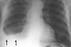
| A 45-year-old woman with a recurrent right temporal glioblastoma multiforme shown on T1-weighted gadolinium-enhanced (T1Gd) MRI. Note the much more prominent tumor to gray matter (T/G) delineation at the later time point, 473 min compared to 90 min. Reprinted by permission of the Society of Nuclear Medicine from Spence AM, Muzi M, Mankoff DA, et al, "18-FDG PET of Gliomas at Delayed Intervals; Improved Distinction Between Tumor and Normal Gray Matter" (J Nucl Med 2004; 45: 1653-1659, p. 1655 [Figure 2]). |
The patients fasted for a minimum of nine hours before the PET studies were undertaken. The study group had an attenuation scan performed with a rotating sector source of germanium-68 through the brain and tumor-containing region, according to the researchers. They also established an intravenous line for isotope injection, and inserted a wrist radial artery line for plasma sampling for the isotope time-activity curve, the authors reported.
The researchers began acquiring emission tomograms on an Advance PET system (GE Healthcare, Waukesha, WI) one minute before the FDG injection.
"Imaging data were collected and reconstructed in four 20-second, four 40-second, four 60-second, four 180-second, and 14 five-minute time bins," the researchers wrote. "Three consecutive five-minute bins were collected at each later time point investigated."
Their image analysis consisted of selecting regions of interest (ROIs) in the gliomas that included contrast-enhancing volume and adjacent nonenhancing tumor defined on the MR images. The PET scan ROIs were drawn while referencing the MR images, according to the authors.
 |
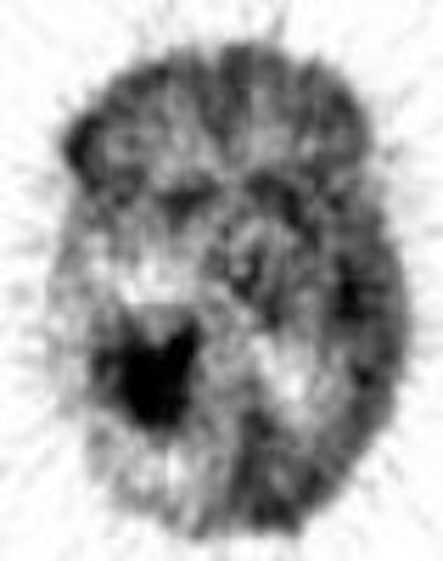
| A 38-year-old man with a recurrent left temporal astrocytoma. Coregistration of T1Gd MRI planes with the F-18 FDG PET planes and use of delayed imaging made evident that the small focus seen on MRI was a tumor rather than radionecrosis, especially at 415 min. Subsequent MRI confirmed rapidly progressing disease. Reprinted by permission of the Society of Nuclear Medicine from Spence AM, Muzi M, Mankoff DA, et al, "18-FDG PET of Gliomas at Delayed Intervals; Improved Distinction Between Tumor and Normal Gray Matter" (J Nucl Med 2004; 45: 1653-1659, p. 1655 [Figure 3]). |
"Brain ROIs were generated on coregistered MR image slices superior to the ventricles and transferred to coordinate F-18 FDG dynamic and static image sets," the researchers wrote.
The authors also analyzed the data by a linear-effects model to determine whether there was a discrete time threshold after which further delaying imaging added no further clinically relevant information. According to the researchers, the intent was to define an optimal delayed time point later than the standard that could be efficiently used clinically and still improve delineation of a tumor from normal brain reference regions.
In 12 of the19 patients, the researchers found that visual analysis of the delayed images (greater than 200 minutes) showed better delineation of the high FDG uptake in the tumors relative to gray matter than standard FDG imaging times of 30-90 minutes. They also found that standardized uptake values (SUV) showed greater uptake in the tumors relative to gray matter, whole brain, or white matter at the delayed times, and that all ratios increased by roughly 20% between the early and late PET imaging times.
The authors noted that their methodology of delaying PET acquisition showed better delineation from brain gray matter for the high-grade (nine of 11 patients) and low-grade tumors (three of five patients), but it did not occur in the stable low-grade tumors (zero of three). In addition, their effort to determine a discrete time threshold for delayed PET imaging proved problematic.
"Our data analysis to determine a time point after which longer delayed imaging added no further clinically relevant information did not define a discreet time threshold," the researchers wrote. "The longer the image was delayed within the time limits we studied; the better was the delineation, except for the three stable low-grade cases."
By Jonathan S. Batchelor
AuntMinnie.com staff writer
November 5, 2004
Related Reading
New study looks into brains of Alzheimer's patients, October 14, 2004
Changing market for PET brings challenges and opportunities, September 30, 2004
CMS to cover PET for Alzheimer's, September 17, 2004
Combined PET and MRI distinguish necrosis from tumor recurrence, October 29, 2002
MRI and SPECT garner high marks for glioma grading, March 5, 2001
Copyright © 2004 AuntMinnie.com