Three-dimensional ultrasound offers more than an easy and accurate way to diagnose conditions of the female reproductive system. It's an opportunity for sonography to reassert itself in radiology by delivering the kinds of 3D insights previously limited to other modalities.
That's the message delivered by Dr. Beryl Benacerraf, a leading researcher and clinical professor of radiology and obstetrics and gynecology at Harvard Medical School in Boston. Now that 3D technology provides ultrasound with its own reconstructed views, she said, sonography can attract more of the interest that has gone to CT and MR in recent years.
"I think this is going to put ultrasound back on the map of cross-sectional imaging," Benacerraf stated in a presentation at the 2004 American Institute of Ultrasound in Medicine (AIUM) conference in Phoenix. "This provides many more opportunities for ultrasound than we have ever had before."
"3D is, in my opinion, one of the most important advances in modern sonography," she continued. "And it is going to explode from here on in, because we will see an enormous amount of development of this technology."
Useful in practice
In her talk, Benacerraf described many specific, often very practical benefits she has found in using 3D ultrasound for pelvic imaging.
For one thing, 3D lends a forgiving attitude to a modality that is notoriously operator-dependent. No longer does the ultrasound image depend on the precise probe placement: the 3D technology obtains a volume scan from which any needed view can be selected.
"You don't have to lament that the patient is gone, or yell at your resident or sonographer who didn't take the right picture. You can just go back to your volume and reconstruct it," Benacerraf noted.
 |
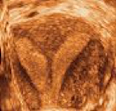
| Three-dimensional ultrasound images showing septate uteri. Images courtesy of Dr. Beryl Benacerraf. |
 |
The volume data generated by 3D technology can also be acquired in less than a minute with just a few sweeps of the probe. While a physician might ordinarily choose to spend more time with the patient obtaining the best views, and less time reconstructing at the computer, the speed of 3D becomes very helpful in procedures such as sonohysterography.
"Taking one picture at a time on these cases is a real drudgery for both you and the patient," Benacerraf said. "But even if you can just take one or two sweeps very quickly, I guarantee you won't have to put in that much fluid because, as you squirt your fluid, you've got your volume. You can reconstruct it, and navigate through it long after the patient's gone."
Another advantage of 3D reconstructions is they are more easily downloaded and forwarded to referring physicians than traditional video clips of ultrasound exams. And they demand far less interpretive skill from the receiving physician than 2D images, Benacerraf said.
Diagnostic distinctions
The pelvic area reconstructions enabled by 3D also provide physicians the views they were denied in the standard image-acquisition planes, but which are, interestingly, more akin to those seen in standard medical illustrations of the female reproductive anatomy, Benacerraf said.
Many of the reconstructed images also make a difference in diagnosis, she said.
Foremost among these is the coronal view of the uterus, an only-in-3D image that can much more definitively distinguish the bicornuate from the septate from the unicornuate uterus.
 |
| Three-dimensional ultrasound images showing submucosal fibroids. Images courtesy of Dr. Beryl Benacerraf. |
 |
 |
"These kinds of images are incredibly important, particularly for the infertility patient," said Benacerraf, noting that women with septate uteri are at much higher risk for miscarriage, preterm labor, and other adverse effects.
With 3D, the sonographer can also measure the width and the depth of the septum, Benacerraf said, "which will be a very good guide for the surgeon who is going to be resecting the septum."
Other instances in which 3D is helpful include identifying the presence and nature of submucosal fibroids, effectively performing a "virtual hysteroscopy."
In one study of 43 patients with abnormal uterine bleeding, Benacerraf and colleagues found that a 3D coronal view of the uterus was helpful in detecting polyps or fibroids in 35% of cases. The polyps were suggested but ill-defined on 2D ultrasound, she said, and 3D clarified the findings.
The same study also found that turning on the 3D capability wasn't necessary or worthwhile if the 2D scan appeared normal or showed a well-defined finding.
Three-dimensional ultrasound is also useful for distinguishing hydrosalpinx and septate cysts in the uterine adnexa, and for imaging the perineum in general, Benacerraf said.
Overall, she concluded, 3D ultrasound is poised to completely and permanently change the views in pelvic imaging. "(In) five to 10 years we will reminisce about viewing images only in the acquisition planes," Benacerraf predicted.
And, if practitioners bring new energy to the field, 3D could also enable ultrasound to challenge the ascendancy of MRI and CT for many imaging needs, she said.
"It is really up to the ultrasound community to work on this, and to demonstrate how we should scan the pelvis. Perhaps we can get a lot more information than we've ever had the opportunity to get using ultrasound," Benacerraf said. "And this will cement ultrasound's role in cross-sectional imaging in this area."
By Tracie L. Thompson
AuntMinnie.com staff writer
September 9, 2004
Related Reading
Sonographic sign reliably excludes ectopic pregnancy, June 22, 2004
Study outlines sonographic findings of pelvic congestion syndrome, April 14, 2004
3-D and the future of imaging, November 3, 2003
3D ultrasound offers reliable assessment of gallbladder, bile ducts, May 5, 2003
3-D ultrasound requires understanding of artifacts, July 8, 2002
3-D ultrasound excels at detecting ovarian cancer, March 14, 2001
Copyright © 2004 AuntMinnie.com