A comprehensive stress MRI exam is more sensitive than stress echocardiography in detecting coronary artery disease in patients who arrive at an emergency room with chest pain, according to a newly reported pilot study.
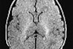
Researchers at Duke University in Durham, NC, looked at the real-world performance of a comprehensive stress exam with rest and adenosine stress first-pass perfusion MRI (pMRI), cine and contrast-enhanced MRI (CE-MRI). They presented their findings at the 2004 American College of Cardiology meeting in New Orleans.
"There is a lot of literature right now on stress perfusion (imaging), but most of it is quantitative. We found that to be quite laborious and difficult to do in an active clinical service like we have at Duke," said Dr. John Heitner, a fellow at the Duke Cardiovascular Magnetic Resonance Center and lead researcher on the study.
"We wanted to really assess stress and MRI perfusion more on a visual assessment, which is what you would do in everyday clinical practice," Heitner said.
The study compared stress MRI with the stress echocardiography exams used as the standard of care at Duke. The pMRI exams were performed using SR GRE-EPI (4-5 short-axis slices/RR, 0.0625 mmol/kg of gadolinium contrast), and interpreted qualitatively with cine and CE-MRI.
Researchers enrolled 60 patients who presented at the emergency room with chest pain and no history of coronary artery disease. Of these, 16 patients who had either a positive stress echo or MRI exam subsequently underwent coronary angiography.
Patients were considered true-positive for coronary artery disease if they had greater than 50% stenosis at angiography or had experienced a clinical event (death, myocardial infarction, or unstable angina) prior to follow-up 7.5 months later.
True-negatives were defined as those with 50% or less stenosis at angiography, or negative stress echo and MRI and no clinical events at follow-up.
None of the patients discharged from the emergency department had experienced a clinical event as of follow-up. Of the 16 patients who underwent coronary angiography, eight were true-positive for coronary artery disease.
However, five patients who would have been discharged from the emergency department based on the negative stress-echo interpretation turned out to have 95% or greater stenosis in a major epicardial vessel.
In clinical interpretations, the stress MRI performed with 100% sensitivity and 90% specificity, compared to echo's 38% sensitivity and 96% specificity. Heitner cautioned that there is a learning curve with stress MRI due to the frequency of imaging artifacts.
And although his study covered only a few patients, Heitner believes stress MRI will become much more commonly used, as it already has by Duke's emergency room.
"The take-home message is that stress-perfusion MRI can be done quickly, effectively, and quite accurately in the clinical setting," Heitner said.
"I think that as the data accumulates, we will see that stress MRI has the sensitivity of stress nuclear and the specificity of stress echo," Heitner said. "With the other advantage that it offers, as far as looking at the aorta and viability, you're going to find it used in most cardiology practices."
By Jerry IngramAuntMinnie.com contributing writer
May 26, 2004
Related Reading
Accuracy of multidetector CT for coronary stenosis depends on patient selection, March 15, 2004
Cardiac MRI tracks impact of statin therapy on plaque, March 7, 2004
Myocardial perfusion measured by MRI useful in detecting heart disease, August 4, 2003
Definitive diagnostic testing useful in ER patients with non-MI chest pain , July 21, 2003
Copyright © 2004 AuntMinnie.com