CT body composition assessment shows that asymptomatic adults with a high accumulation of muscle fat -- a condition called myosteatosis -- are at increased risk of major adverse cardiovascular events, according to a study published on May 16 in Radiology.
The study findings could help clinicians better understand the role myosteatosis can play in a patient's health, wrote a team led by Dr. Maxime Nachit, PhD, of the University of Louvain in Brussels.
"To date, most of the medical interest on body composition and its impact on health is directed toward (visceral) obesity, liver steatosis, and muscle wasting, while myosteatosis is overlooked," the group noted. "[But] myosteatosis robustly associates with prognosis in patients with chronic diseases."
Body composition tends to be assessed using the body mass index, or BMI, which is calculated using a person's height and weight, and most assessments focus on visceral fat and liver steatosis. Myosteatosis is fat that accumulates in the muscles, and it tends to be identified via CT or MR imaging in people who are already sick and being treated for another condition. How myosteatosis affects asymptomatic individuals' health has been underexplored, Nachit and colleagues wrote.
"The typically silent accumulation of fat in ectopic locations is now considered to be one of the major risk factors of mortality, independent of obesity," the team noted.
Nachit's team sought to evaluate any links between myosteatosis, adverse events (such as heart failure, heart attack, or aortic aneurysm) and mortality risk via a study that included data from 8,982 adults. Patients underwent low-dose unenhanced abdominal CT imaging for colorectal cancer screening between April 2004 and December 2016; the images were processed with an artificial intelligence (AI)-based body composition algorithm which tracked connections between obesity (2,994 patients), liver steatosis (4,395 patients), myopenia (1,589 patients), myosteatosis (2,245 patients), and risk of mortality.
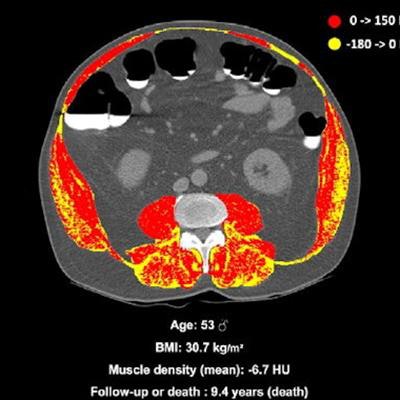
 (A) Unenhanced axial abdominal CT image with a Hounsfield unit-based color scale of skeletal muscles in a 51-year-old man with obesity, smoking history, no type 2 diabetes, and no history of cardiovascular events at inclusion shows mild fatty infiltration in the muscles (myosteatosis, yellow), with most voxels in the positive range of Hounsfield units (red). The patient was lost to follow-up after 13.2 years. (B) Unenhanced axial abdominal CT image with a Hounsfield unit–based color scale of skeletal muscles in a 53-year-old man with obesity, smoking history, no type 2 diabetes, and no history of cardiovascular events at inclusion shows severe fatty infiltration in the muscles (myosteatosis, yellow), mostly distributed in the paravertebral (i.e., erector spinae and multifidus) and oblique muscle groups. The patient died after 9.4 years of follow-up. Image and caption courtesy of the RSNA.
Of the all the study participants, 507 died over the course of three years of follow-up after baseline imaging. The group found myosteatosis in 55% of study participants who died; absolute mortality risk at 10 years in persons with myosteatosis was 15.5% compared to obesity (7.6%), liver steatosis (8.5%), or myopenia (9.7%).
Although health factors such as visceral fat and liver steatosis were also associated with higher mortality risk, myosteatosis remained the highest, the team reported. It also found that the mortality risk of individuals with myosteatosis was comparable to that associated with smoking or having type 2 diabetes, according to the authors.
| Overall mortality risk in asymptomatic patients screened for colon cancer | ||
| Parameter | Hazard ratio (1 as reference) | p-value |
| Myosteatosis | 1.89 | < 0.001 |
| Type 2 diabetes | 1.56 | 0.02 |
| Myopenia | 1.38 | 0.008 |
| Liver steatosis | 1.32 | 0.006 |
| Body mass index | 1.01 | 0.6 |
| Visceral fat | 1 | 0.7 |
| Male sex | 0.96 | 0.7 |
"[The] relationship [between myosteatosis and mortality risk] was independent from age or markers of obesity such as BMI," Nachit said in a statement released by the RSNA.
The study findings could help clinicians further customize patients' health assessments, Nachit said.
"We are witnessing the onset of 'personalized medicine,' whose aim is to tailor medical management at the individual level based on a constellation of information such as genetics, medical history, physical characteristics, complex and large-scale molecular evaluation," she said.
The research highlights how regular screening -- in this case, for colorectal cancer -- can have a beneficial effect on patients' health, according to Dr. Angela Tong of New York University and Dr. Kirti Magudia, PhD, of Duke University in Durham, NC.
"Opportunistic screening of body composition from abdominal CT has the potential to impact overall health care by extracting latent value from CT being performed for other indications," Tong and Magudia noted in an accompanying editorial.