A commercially available artificial intelligence (AI) program designed to help interpret patient chest x-rays shows promise for shortening overall reading times for radiologists -- although not when it detects abnormalities, according to a study published April 29 in npj Digital Medicine.
A group at Yonsei University in Seoul, South Korea, calculated reading times for radiologists who interpreted a minimum of 300 chest x-rays per month over a two-month period, either with or without the aid of an AI program (Insight CXR, v3, Lunit). The team found that using AI reduces the time radiologists spend interpreting images that turn out to be normal, first author Dr. Hyun Joo Shin, PhD, and colleagues wrote.
"Overall reading times shortened when radiologists referred to AI, especially for normal [chest x-rays]; however, abnormalities detected by AI on [chest x-rays] appeared to lengthen reading times," the group noted.
The researchers have been assessing the value of the software in studies since they integrated it at their hospital in March 2020. In this latest research, they evaluated how the AI program might affect radiologists' workload.
Between September and December 2021, the group assessed reading times of 11 radiologists who interpreted chest x-rays for two months using the software and for two months without it. Reading time was defined as the duration in seconds from opening chest x-rays to filing a report on the image in their PACS. A total of 18,680 images were included in the study.
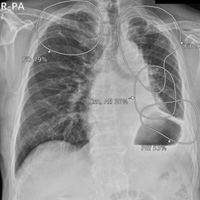
 Integration of AI for chest x-rays on PACS. (a) The AI result attached to the second image of the original chest x-ray contains a contour map, abbreviations, and the abnormality score of detected lesions. Doctors can simply refer to the AI results by scrolling down the original image on the PACS. (b) The highest abnormality score is used as the total abnormality score of each image, and this was listed as a separate column (red square) on the PACS. Image and caption courtesy of npj Digital Medicine through CC BY 4.0.
The investigators compared reading times according to the presence of lesions detected by AI for any one of the following eight abnormalities: atelectasis, cardiomegaly, consolidation, fibrosis, nodule, pleural effusion, pneumoperitoneum, or pneumothorax.
Total reading times were significantly shortened with AI use, compared to no use, and when there were no abnormalities, reading times were shorter with AI use.
| Reading times for chest x-rays, with and without AI support | |||
| Measure | Without AI support | With AI support | p-value |
| Total reading time | 14.8 seconds | 13.3 seconds | < 0.001 |
| Reading time for normal x-rays | Mean, 13.1 seconds | Mean, 10.8 seconds | < 0.001 |
But if AI detected any abnormality, reading times did not differ according to AI use (mean 18.6 seconds vs. 18.4 seconds, p = 0.452). In fact, reading times increased as abnormality scores increased, the researchers reported.
The finding that reading times increased as abnormality scores increased could be due to radiologists taking more time to judge the validity of the AI assessment and to report more details about the findings seen on images, regardless of the accuracy of displayed AI results, the researchers suggested.
Nonetheless, the findings are positive overall, according to the researchers.
"AI may be able to improve the efficiency of radiologists by sparing time spent on normal images and allowing them to invest this time in [chest x-rays] with abnormalities," they concluded.