Dynamic digital radiography (DDR) estimates for measuring blood flow in the lungs matched lung perfusion scintigraphy in a group of lung transplant patients, according to research presented at the American Roentgen Ray Society (ARRS) annual meeting in Honolulu.
In a session held April 19 on cardiac and chest imaging, Dr. Padma Manapragada of the University of Alabama Birmingham presented a study comparing DDR with lung scintigraphy perfusion exams and found the two exams provided almost equal diagnostic information. The finding bodes well for further development of DDR, she said.
"DDR estimates for differential lung perfusion are strongly correlated with results obtained by [nuclear medicine] lung scintigraphy," Manapragada said.
Lung scintigraphy exams capture images of blood flow in the lungs based on the distribution of an injected radiotracer. These standard nuclear-medicine procedures can provide critical information for clinicians when diagnosing pulmonary embolism, for instance.
Conversely, DDR is a relatively new technique based on conventional x-ray, but that uses a pulsed generator and flat-panel digital detector to acquire images at 15 frames per second for cardiopulmonary applications. Once reconstructed by computer software, DDR images reveal the lungs in motion. These anatomical views may provide clinicians with more functional information than standard imaging, researchers suggest.
In this pilot study, Manapragada and colleagues compared the two approaches in a group of 29 preoperative lung transplant patients who underwent both tests at their hospital between January and October 2022. They calculated lung perfusion as percentages for both tests and then compared them.
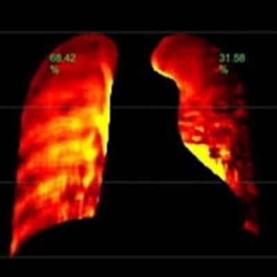
 Dr. Padma Manapragada of the University of Alabama Birmingham presented a study of the use of dynamic digital radiography in preoperative lung transplant patients at ARRS 2023 in Honolulu. Image courtesy of Dr. Manapragada.
"There were no discrepancies between the two modalities in determining which lung was better perfused," Manapragada said.
Specifically, the analysis revealed a strong statistical match between the perfusion results obtained using the two approaches, with a Pearson's correlation coefficient of 0.945, according to the findings.
In one case Manapragada presented, preoperative scintigraphy perfusion estimates were 35% in the left lung and 65% in the right lung, while DDR perfusion estimates were 32% in the left lung and 68% in the patient's right lung, she noted.
Ultimately, this was a small pilot study, and further work is needed to confirm the correlations between lung scintigraphy and DDR, Manapragada said. Specifically, investigators plan to look at whether patient positioning affects results, as patients in this study were standing during the DDR exam and supine for perfusion scintigraphy, she noted.
"If these results are confirmed, DDR has the potential to be a valuable adjunct or possible replacement for nuclear medicine lung scintigraphy," Manapragada concluded.