FDG-PET/CT imaging shows metabolic neurologic changes due to COVID-19 in patients before the onset of anatomical abnormalities and can detect more active infection sites than MRI, according to research presented March 2 at ECR 2023 in Vienna.
In a research session covering central nervous system infection, Dr. Nikhil Seniaray of Mahajan Imaging in New Delhi, India, presented a study that looked at brain imaging findings using the two approaches in patients diagnosed with COVID-19 three months before the appearance of neurological symptoms.
"FDG-PET/CT may help in certain acute and subacute neurological manifestations of COVID-19, especially with noncontributory MRI, thereby expediting subsequent clinical management," Seniaray said.
COVID-19 is now universally accepted as a disease affecting almost all organ systems in the body, with CNS involvement seen in up to 4% of all patients, Seniaray explained.
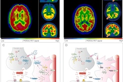
While MRI is the primary tool in cases of suspected neurological complications, preliminary research suggests that FDG-PET/CT scans may show additional areas of involvement in the brain in the form of hypometabolism or hypermetabolism as a result of synaptic dysfunction, he said.
 Dr. Nikhil Seniaray of Mahajan Imaging in New Delhi, India, presented a study March 2 that looked at brain imaging findings in patients with COVID-19.
To elucidate these findings further, the researchers performed a retrospective study in 30 patients with COVID-19 infection who had undergone imaging within three months of the appearance of symptoms. Based on MRI scans, patients were classified as positive (n = 14) and negative (n = 16).
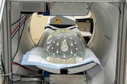
Patients also underwent either total-body PET/CT (uMI 550, United Imaging) or whole-body PET/CT scans (Discovery STE, GE HealthCare) following injections with F-18 FDG radiotracer and a waiting period of 60 minutes. Patients were scanned in one bed position for 10 minutes.
Out of the 16 MRI-negative patients, nine had clusters of hypermetabolism in the frontal, insular, and cingulate cortices brain regions, according to the findings, and seven showed posterior cortical-subcortical and cerebellar hypermetabolism on FDG-PET/CT images. Antibodies to COVID-19 were confirmed in serum of 28 patients and in cerebral spine fluid in five patients, Seniaray added.
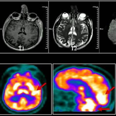
In a presentation of cases, Seniaray showed images of a 43-year-old patient who reported a brief history of febrile illness with seizures and limb weakness. The MRI scan was noncontributory, while the PET/CT scan showed increased F-18 FDG radiotracer uptake in the bilateral cerebral and cerebellar brain hemispheres, likely inflammatory, he said.
Moreover, the patient was put on immunomodulatory treatment and a follow-up FDG PET/CT scan one week later showed resolution of the hypermetabolism, he said.
Ultimately, the study supports published literature suggesting neurological manifestations of COVID-19 can be seen even in patients with mild to moderate disease and that FDG-PET/CT can be a useful tool for assessing patient response to therapy, Seniaray said.
"FDG PET/CT can detect active functional or metabolic changes before the onset of anatomical abnormalities and can detect more active infection sites as compared to MRI," he concluded.