MRI-identified bone markers -- such as porosity, morphologic structure, mineralization, and density -- are useful for assessing bone health in postmenopausal women, a study published January 24 in Radiology has found.
The findings could translate into better patient care for postmenopausal women with osteoporosis, noted a team led by Dr. Brandon Jones of the University of Pennsylvania in Philadelphia.
"In this preliminary study in postmenopausal women, MRI markers helped detect impairments in cortical bone quality associated with osteoporosis, and all markers were associated with bone mineral density," the investigators wrote.
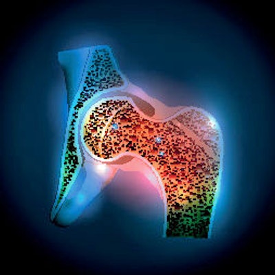
Osteoporosis is associated with two million annual fractures and more than $57 billion in healthcare costs, the group explained. Risk of osteoporosis-related fractures is typically predicted by bone mineral density assessment using dual-energy x-ray absorptiometry (DEXA), but although this technique measures density it cannot distinguish between trabecular and cortical bone -- the health of which influences fracture risk.
Measuring cortical bone health, particularly porosity, has been linked to better prediction of fracture risk; this has tended to be done with CT imaging. But MRI has become a more viable alternative.
"Advances in solid-state MRI techniques of ultrashort echo time ... and zero echo time sequences [have] enabled quantification of previously undetectable signals in tissues with solid-like MRI properties," the group wrote.
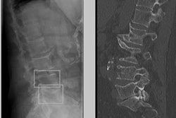
The team sought to explore whether MRI markers of bone health could help predict fracture risk in postmenopausal women with osteoporosis. The group conducted a study that included 34 postmenopausal women recruited between January 2019 and October 2020. Of these, 15 had osteoporosis (identified on dual-energy x-ray absorptiometry, or DEXA) and had not undergone any treatment for it, and 19 did not have the disease.
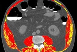
All underwent MRI and CT in the mid-tibia and DEXA in the hip and spine. The investigators specifically tracked MRI measures of bone porosity, density, and mineralization, comparing these results among the osteoporosis and non-osteoporosis groups.
Overall, the women with osteoporosis had higher bone porosity and lower cortical bone thickness and mineralization, the group found.
| MRI findings regarding bone health in women with and without osteoporosis | |||
| Measure | Women without osteoporosis | Women with osteoporosis | p-value |
| Bone porosity (measured as elevated pore water) | 9.5 mol/L | 11.6 mol/L | 0.007 |
| Cortical bone thickness | 5.6 mm | 4.8 mm | 0.001 |
| Mineralization (measured as phosphorus density) | 7.5 mol/L | 6.4 mol/L | 0.01 |
The study findings show promise, but more research is indicated, according to the team.
"Solid-state MRI was able to help detect impairment in cortical bone porosity, morphologic structure, and mineralization in participants with postmenopausal osteoporosis" it concluded. "Future work should investigate these markers in larger groups to determine if they can detect changes in bone health following pharmacologic treatment."