Exposure to opioids in utero has a negative effect on fetal brain development, a study published January 18 in the American Journal of Roentgenology indicates.
The results could help clarify best postnatal care for infants exposed to opioids, wrote a team led by Dr. Usha Nagaraj of Cincinnati Children's Hospital Medical Center in Ohio.
"A full understanding of how opioids affect the developing brain ... requires prenatal studies that evaluate the brain before the effects of opioid withdrawal and other postnatal variables (e.g., type of infant feeding, neonatal intensive care unit stay, and maternal infant bonding) can be assessed," the group noted.
More than 100 people die of opioid overdose in the U.S. each day, and previous research has shown that infants experience consequences of adult opioid use, tending to have lower birth weights, smaller head circumferences, increased white matter injury, and altered functional networks, Nagaraj and colleagues noted. But not as much is known about the effect of opioids on fetuses.
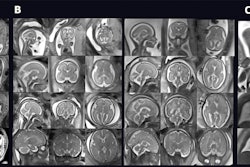
To address this knowledge gap, Nagaraj's group conducted a study that included 65 women in their third trimester of pregnancy who underwent fetal MRI exams between July 2020 and December 2021. Of the 65 fetuses, 28 had been exposed to opioids. The investigators tracked 14 biometrics of fetuses' brains and adjusted the results for gestational age, fetal sex, and exposure to nicotine.
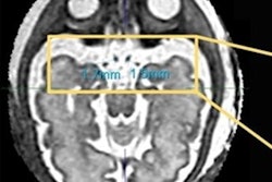
Of the 14 brain biometrics the group measured, seven were smaller in fetuses with opioid exposure compared with unexposed counterparts, suggesting negatively altered physiology due to the drug. With the exception of two measurements, all were statistically significant.
| Comparison of brain biometric measurements in fetuses not exposed and exposed to opioids | |||
| Brain metric | Not exposed | Exposed | p-value |
| Cerebral fronto-occipital diameter | 95 mm | 93.8 mm | 0.03 |
| Bone biparietal diameter | 80.3 mm | 79 mm | 0.01 |
| Brain biparietal diameter | 74.1 mm | 72.9 mm | 0.49 |
| Corpus callosum length | 39.4 mm | 37.7 mm | 0.45 |
| Vermis height | 18.8 mm | 18.2 mm | 0.002 |
| Anteroposterior pons measurement | 12.1 mm | 11.6 mm | 0.006 |
| Transverse cerebellar diameter | 41.4 mm | 40.4 mm | 0.02 |
When the group adjusted the study results, it found that exposed fetuses were more likely to be in the breech position (21% compared with 3%) and to have increased amniotic fluid volume (29% compared with 8%). The cause of these conditions in relation to opioid exposure was unclear, but the authors did note that in particular, breech presentation is associated with "higher risk of congenital anomalies" and obstetric risk factors such as fetal growth restriction and gestational diabetes.
The investigators conceded that the study had some limitations, including a small sample size, potentially confounding fetal exposure to nicotine or other drugs as well as opioids, and the fact that the brain development of the fetuses could also have been affected by maternal nutrition or stress. But the results do shed light on how opioid exposure affects infants in utero.
"[Our] findings provide insight into the impact of prenatal opioid exposure on fetal brain development," the group concluded.