Commercially available software designed to enhance the quality of digital x-rays appears to be a feasible way to visualize whether chest tubes, catheters, or wires are placed correctly in patients, according to a study published November 14 in Radiography.
A group led by Helle Precht, PhD, of the University College Lillebaelt in Odense explored for the first time whether Canon Medical Systems Europe's Advanced Edge Enhancement (AEE) software could be clinically useful for this purpose.
"The use of AEE algorithms has the potential to reduce the number of retakes in this patient group, by improving visualization of tubes, catheters, and wires on initial radiographs, rather than requiring modification of exposure parameters and irradiation of the patient," the group wrote.
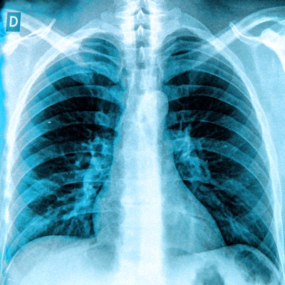
X-ray is the standard approach for determining the correct placement of devices in patients' chests, as incorrect placement can lead to complications, such as pleural effusion, pulmonary edema, chest wall abscess, and chest or back pain.
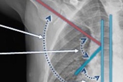
The AEE software is installed in Canon's digital x-ray machines and applies three different user algorithms (catheter, bone, and small structure) to enhance the visualization of the edges of these targets from surrounding tissue. While the software has been shown to improve visual characteristics in other anatomical areas, to date, no studies have explored its usefulness in chest x-rays, according to the authors.
The researchers included 50 consecutive anterior-posterior chest x-rays acquired from patients with either a tube, catheter, or wire present. All three of the software's algorithms were applied to the chest x-rays in postprocessing using two intensity settings (1 and 4), for a total of 350 different images. Three radiologists then evaluated the images using a subjective grading analysis; a score of three indicated they were suitable for diagnostic assessments.
According to the findings, the three AEE algorithms contributed to an overall improvement (average range between 16% to 49%) in the visualization of tubes, catheters, or wires on the chest x-ray images. It also demonstrated a statistically significant (p < 0.05) improvement in contrast resolution and sharpness.
However, the high-intensity catheter algorithm was the only one to achieve a statistically significant (p = 0.017) increase in the ability to differentiate pulmonary tissues of similar density, according to the authors.
"The bone and catheter algorithms showed the highest consistency, with the small structure algorithm underperforming in resolution and low contrast resolution," they wrote.
While the results are encouraging for more clinical adoption of the software, future studies are needed to further investigate which AEE settings are the most effective for different patient sizes, medical devices, sexes, and patient positioning, the authors wrote.
"The algorithm should also be evaluated in clinical practice to measure its effect during a clinical workday," Precht and colleagues concluded.