In this poster session, Stanford researchers will share results from a literature review that consisted of 11 studies gleaned in April of this year from Medline, Embase, and Cochrane databases that addressed the use of chest MRI to identify lung nodules equal to or greater than 4 mm that are suspicious for malignancy. The studies included data from 963 patients and 1,149 nodules and used chest CT as the performance comparison standard. The investigators compared the two modalities' per-lesion and per-patient sensitivity and specificity.
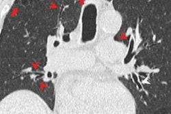
What did the study find? Per-lesion sensitivity of MRI was 88% for detecting suspicious lung nodules between 4 mm and 30 mm; its sensitivity for nodules less than 10 mm was 85% and its sensitivity for nodules 10 mm to 30 mm was 99%. Finally, MRI's per-patient specificity was 94%.
"Chest MRI can be a reasonable alternative to CT in the detection and follow-up of pulmonary nodules equal to or less than 4 mm in patients with a high radiation burden," the team concluded in its abstract.
Learn more by sitting in on this presentation.