PET imaging may be a useful tool for guiding and monitoring treatment success in prostate cancer patients undergoing high-intensity focused ultrasound (HIFU), according to a study published November 2 in the Journal of Nuclear Medicine.
A team led by Dr. Andrei Iagaru of Stanford University evaluated whether using both gallium-68 (Ga-68) RM2 and Ga-68 PSMA-11 PET imaging radiotracers in prostate cancer patients before and after HIFU could determine their response to the treatment. Early results are promising, the group found.
"Focal therapy for localized prostate cancer using HIFU is gaining in popularity as it is noninvasive and associated with fewer side effects than standard whole-gland treatments," Iagaru and colleagues wrote. "However, better methods to evaluate response to HIFU ablation are an unmet need."
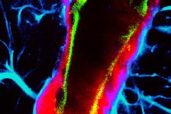
Prostate-specific membrane antigen (PSMA) and gastrin-releasing peptide receptors (GRPR) are molecules that are overexpressed by prostate cancer cells and are believed to contribute to the growth of tumors. Ga-68 RM2 and Ga-68 PSMA-11 are radiotracers designed to bind to these targets, and thus reveal the extent of the cancer of PET scans.
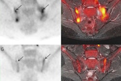
The researchers retrospectively enrolled 14 patients between the ages of 48 and 78 with newly diagnosed prostate cancer who were scheduled for HIFU treatment. Patients underwent Ga-68 PSMA-11 PET/MRI scans followed by Ga-68 RM2 PET/MRI within two weeks, or vice versa, and then again at least six months after HIFU.
Treatment response was measured by comparing uptake levels of the radiotracer by the tumors, specifically maximum standardized uptake values (SUVmax). To establish a benchmark, patients also underwent MRI and biopsies to locate and confirm lesions.
Pre-HIFU, Ga-68 PSMA-11 identified all target tumors (23), while Ga-68 RM2 missed two. Post-HIFU, both tracers identified clinically significant residual disease in one patient. In addition, pretreatment prostate-specific antigen decreased by 66% after HIFU, and this decrease correlated with a significant drop in SUVmax after HIFU.
"Ga-68 PSMA-11 and Ga-68 RM2 PET/MRI identified the target tumor for HIFU in 100% and 86%, respectively, and accurately verified response to treatment," the authors wrote.
Ultimately, this was a pilot study and although the results are promising, more work is needed, the authors noted.
"PET might be a useful tool in the guidance and monitoring of treatment success in patients receiving focal therapy for prostate cancer. These preliminary findings warrant larger studies for validation," the group concluded.