With healthcare advancements, more patient data exists than ever before. But much of that data, including radiology images and pathology scans, is housed in disparate systems that make it difficult for physicians to access the information necessary to obtain a holistic view of a patient's health.
 Jonathan Shoemaker.
One health system is trying to change that. Led by Jonathan Shoemaker, the imaging systems team at Stanford Health Care in California has spent the past several years developing an enterprise imaging system that will integrate pathology.
Shoemaker, who serves as administrative director of imaging systems and services, said that the goal is to make it easier for physicians to correlate imaging data across medical specialties so that they can form a more comprehensive understanding of individual patients and better track conditions over time.
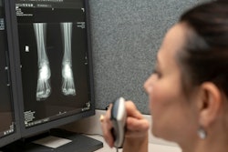
"Typically, images are stored within silos within the organization, and they're not easily accessible by all physicians or even patients themselves," Shoemaker said. "But a picture's worth 1,000 words. While the report may be descriptive, and the reports and results are available throughout the organization to other physicians, that's not always enough. If you're looking at tumor size progression, for example, it's always good to be able to compare the new imaging to the prior exam, and that's difficult to do if you can't access the previous studies."
Creating a complete medical record
Stanford began exploring enterprise imaging when it came time to replace its PACS. Rather than swapping its PACS with another system dedicated solely to radiology studies, the team decided to add enterprise imaging to enhance data sharing among medical specialties.
With enterprise imaging, "you get a more comprehensive 360° review of the patient and a more complete medical record," Shoemaker said. "You also have the ability to collaborate more deeply and broadly once you have all of the images into a single platform. And if a patient wishes to get a copy of their images, you can query them and place them on the same media or share them through a standard mechanism, like an image exchange."
Implementing the system at Stanford has been an iterative process.
"The initial push for the enterprise platform was related to core imaging, which involved radiology, the vendor-neutral archive and enterprise viewer, with normalization of data through the standard data models and other standard data flow into the imaging platform," Shoemaker explained. "We have since added imaging from other specialties. Right now, we have ophthalmology, dermatology, radiology, [ear, nose and throat], functional MRI, and dual-source CT."
And soon they will add pathology. Given that pathology, like radiology, is critical to nearly all diagnoses, integrating it into the system has been a particular priority of the project.
"It creates a better care continuum," Shoemaker said. "If a radiologist makes a diagnosis, that diagnosis is validated with pathology, and being able to route that positive diagnostic perspective back to the performing radiologist closes that loop. If it doesn't match, then it informs the radiologist, and that's something that can be tracked and measured."
DICOM conversion
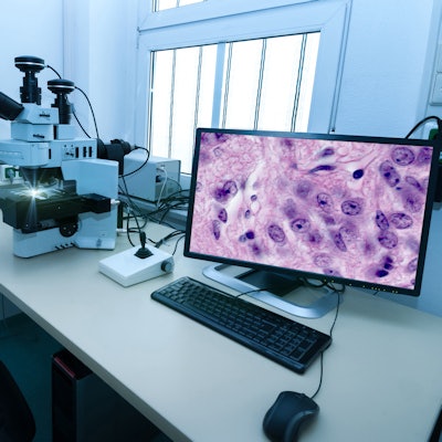
But integrating pathology into the enterprise imaging system hasn't been simple. Although the whole-slide images were integrated into the system using links and a third-party launch, the non-whole-slide images had to be converted into the DICOM standard, which enables healthcare professionals to view, store, and share medical imaging.
"For nonwhole slide images, we are acquiring the data through multiple different types of interfaces, and we are mapping them against the orders and converting the images into DICOM," Shoemaker said.
This is necessary because a lot of pathology devices are not up to date on the standards, he said.
"They don't produce DICOM images, and they don't export DICOM images or utilize the worklist," Shoemaker said. "So, we have to use a third-party tool to basically capture, assign, and wrap those images or convert them and store them for viewing."
The team refrained from using a secondary capture image information object definition (IOD), which converts images from a non-DICOM format to a modality independent DICOM format.
"If you have a bunch of image types of OT [Other], you are not going to know what they are," Shoemaker said. "We've really tried to align a lot of the data to the assigned pathology IOD. For the most part, we've been successful with that. The difficulty that we've had is, obviously, mislabeled data. Data stored using pre-text is always difficult to find the commonality through, and then also mislabeled data within the LIS [laboratory information system] that had to be massaged and placed into the correct type of image, if you will."
The team conducted a proof-of-concept project with 250 non-whole-slide studies to ensure that the data not only converted from the legacy format to DICOM but remained diagnostic in the process with the proper header information to support pathology reading and workflows, according to Shoemaker.
"We had a LIS that acquired and stored the images within its database, and a lot of the formats were open formats, like JPG and TIFF, but all of the metadata associated with those images were archived within that proprietary database," Shoemaker said. "As part of the go-live requirements for our upcoming LIS replacement project, we wanted to migrate those images out. So, we had to work to extract the associated image data, convert the image and write the discrete data into the header information in order for it to not only be sorted but then also be assembled by the platform view in a way that made sense to the pathologist reading the case."
Greater digital advancement
Once the team knew that it could successfully convert the data, it developed an automated process to migrate the pathology images into the enterprise imaging system. That process is underway, and Shoemaker expects the images to be available by mid-November.
"Those will be linked with other image types, like radiology and dermatology, in how we present the data through either an enterprise viewer or a diagnostic viewer," Shoemaker said.
With this phase of the project nearly complete, Shoemaker reflected on the lessons that he and his team have learned. Namely, he said, they have found that while the standards are good, they don't go far enough.
"For instance, if I want to hang a slide of a liver lesion, the body part associated with the slide specimen is not easily aligned with our SNOMED or RadLex body parts," Shoemaker said, adding that a 2021 white paper about body part labeling to enable enterprise imaging helped, but more work needs to be done. "We need the clinical committees and organizations to produce a full molecular to macro body-part tree, and then the standards need to allow for that tree to be cross-walked within the header information, if not better than the header information so that now you could actually display a liver lesion against either a liver ultrasound or an abdomen CT."
Ultimately, Shoemaker hopes that more vendors will create more digital pathology scanners that export images as DICOM to make pathology integration into enterprise imaging easier.
"A lot of scanner vendors are just now coming out with the capability to export as DICOM," he said. "Hopefully that trend will increase with the quality and compressions. Adherence to standards within pathology will only expedite its digital revolution. Some of the algorithms and AI and machine learning that are being produced on the objects themselves are phenomenal, but if you can't run them in flight between the scanner and the solution, it becomes very complex in order to do so in a clinically urgent manner."