PET/CT imaging can predict whether prostate cancer lesions that appear to have spread to patients' bones are malignant or benign, according to a group of researchers at the National Cancer Institute (NCI).
The findings could lead to a reduction in unnecessary biopsies, lead author Tim Phelps, PhD, and colleagues noted. The study results were published October 20 in the Journal of Nuclear Medicine.
"Interpreting such lesions as metastatic without sufficient evidence can have far-reaching implications for patients and lead to unnecessary interventions that alter a patient's quality of life," the group wrote.
Indeterminate bone lesions (IBLs) on PET/CT scans in patients with prostate cancer are common, according to the researchers. Currently, these lesions require biopsy or follow-up imaging for definitive assessment. But these biopsies may not be needed.
Phelps and colleagues explored whether particular features on PET/CT scans performed with F-18 DCFPyL (Pylarify, Lantheus Medical Imaging, approved in the U.S. last year for prostate cancer imaging) could help clinicians determine whether prostate lesions were likely malignant or benign, and decide on effective treatments without the need for biopsy.
The researchers collected data on 243 patients with high-risk primary or biochemically recurrent prostate cancer who showed no prior evidence of metastatic disease. Study participants underwent imaging between July 2017 and October 2021 and were included if they had at least one IBL.
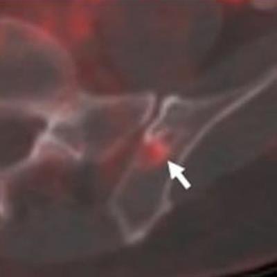
 A 60-year-old patient with biochemically recurrent prostate cancer and a PSA level of 3.9 ng/mL. Axial F-18 DCFPyL PET (left), F-18 DCFPyL PET/CT (middle), and CT (right) images show a single subtle PSMA-avid uptake with SUVmax of 3.9 in the left iliac bone and mixed sclerotic/lytic CT features (arrows). This indeterminate bone lesion (IBL) was stable for four months and negative on four other staging modalities; thus, the IBL was determined to be benign. Image courtesy of the Journal of Nuclear Medicine.
Two nuclear medicine physicians categorized the IBLs on the images as either benign, malignant, or equivocal, noting lesion locations' maximum standard uptake values (SUVmax) for each.
Overall, 98 IBLs were identified in 48 of the 243 patients (19.8%). Of these, 37 were categorized as benign, 42 as malignant, and 19 as equivocal.
Of the benign IBLs, 91.9% showed SUVmax < 5 or exhibited focal uptake without coexisting bone metastases. Conversely, 88.1% of the IBLs deemed malignant demonstrated SUVmax ≥ 5 or were present with coexisting bone metastases.
IBLs on PSMA PET/CT are concerning; however, characterizing their location, SUV, and additional scan findings can aid interpretation," the group wrote.
The study suggests that IBLs are likely malignant in the presence of other bone metastases and that those with SUVmax ≥ 5 at two-hour uptake of F-18 DCFPyL have increased likelihood of malignancy, the authors wrote.
"Understanding specific features that increase the certainty of interpreting IBLs as probably benign or probably malignant is, therefore, of great clinical importance," they concluded.


