Minnies winners, page 3
Scientific Paper of the Year
Minnies 2022 Winner: Mandating Limits on Workload, Duty, and Speed in Radiology. Alexander R, Waite S, et al, Radiology, June 14. Learn more about this study.

The winner selected for Scientific Paper of the Year by the Minnies expert panel recommended hitting the brakes on regulating the workloads of radiologists in response to so-called "reckless reading lawsuits."
Lead author Dr. Stephen Waite of SUNY Downstate Medical Center in Brooklyn, NY, said the idea of the study was to make a scientifically based rebuttal to a 2020 lawsuit in which a plaintiff attorney argued that a radiologist missed a subdural hematoma in a 64-year-old patient because he viewed each image of a CT scan in less than half a second.
The lawyer alleged that the radiologist was lax in his duty and did not spend enough time per image -- and used this argument to negotiate a larger settlement.
"We thought it would be an opportune time to discuss the relationship between speed, accuracy, and fatigue as it relates to performance in radiology," Waite told AuntMinnie.com.
In their review and analysis, the authors focused on the details of the case. The plaintiff's lawyer cited evidence based on subpoenaed keystroke data that the subdural hematoma was missed because the radiologist spent a total of six minutes and 27 seconds reading a CT exam of the head and cervical spine.
By their calculations, this time estimation was simply wrong, the authors found.
In a 2019 study, researchers found that private practice radiologists generate between 13,000 and 15,000 relative value units (RVUs) per year. Given 261 working (nonweekend) days in 2021, and assuming eight weeks of vacation, radiologists would have to generate 63 RVUs per day to achieve 14,000 RVUs per year.
Assuming that a CT scan of the abdomen and pelvis generates 1.82 RVUs, this amounts to approximately 34 scans per day, the authors wrote.
In another study, researchers reported that the average CT exam contains 679 images. By using this figure for the average number of images per exam and a seven-hour workday (assuming a standard eight-hour workday including a one-hour lunch break), this leaves 1.1 seconds for radiologists to look at each image.
Still, this is unrealistic, since it presumes no breaks, interruptions, consultations, or conferences, the authors wrote.
A 2014 study found that on-call radiologists receive an average of 72 phone calls during a typical 12-hour overnight shift, with an average total phone time of 108 minutes. Thus, allowing for 90 minutes of interruptions, breaks, consultations, and conferences, this leaves radiologists with less than one second (0.86 sec) to read each image.
"The available evidence indicates that reading an image in a cross-sectional study in less than a second on average is the current standard of care in radiology, so implying that such behavior is negligent fails logic," the authors wrote.
Robert Alexander, PhD, a co-author and research professor of ophthalmology at SUNY Downstate Medical Center, added that while speed and accuracy are always "connected," the relationship between them is more complex than the legal system is accounting for.
Alexander's work orbits around eye movement analysis in both experimental and applied environments. He studies the relationship between attention, perception, and oculomotor behavior.
"If a radiologist has ten minutes to read an image, but you force them to read it in five minutes, that doesn't mean they will be half as accurate," he told AuntMinnie.com.
The relationship between speed and accuracy is nonlinear, and that nonlinearity depends on many factors, he added. For example, images in a CT stack are not read in the same way as a chest x-ray. They are different beasts, even though both may be images of lungs and both might take up the same size on the reading room's viewing screen. Eye movements and scrolling behavior are very different on a CT than on chest radiographs.
"All of this will change the way you view these different kinds of images. Some individual images in a stack may have a very different viewing time than others, but that does not mean those images were viewed more accurately than the images that were not viewed for as long," he said.
Ultimately, regulating workloads at present with little scientific evidence available could do more harm than good, the authors concluded.
"Although workload limits may ultimately be needed, there is currently no good data from which to set those limits," Waite said. "Without any principled grounding in science, any imposed limits can very likely fail to achieve their desired effect and moreover can lead to a number of unintended consequences ultimately harming patient care."
Runner-up: Reading Race: AI Recognises Patient's Racial Identity In Medical Images. Imon Banerjee I, Bhimireddy A et al, arXiv.org, July 21.
Best New Radiology Device
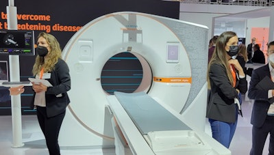
Naeotom Alpha, Siemens Healthineers
Naeotom Alpha made an immediate splash after Siemens Healthineers received U.S. Food and Drug Administration (FDA) clearance in September 2021. The FDA, a government agency certainly not prone to hyperbole, called the proton-counting scanner the first "major imaging device advancement in CT in nearly a decade."
Traditionally, CT scanners have utilized conventional energy integrating detectors, which measure the total energy in many x-rays at once. In photon-counting CT technology as utilized by Naeotom Alpha, each individual x-ray photon is measured as it passes through the patient's body. As a result, photon-counting CT can reduce dose and produce higher-resolution images compared with the conventional approach.
 The Naeotom Alpha CT scanner.
Naeotom Alpha is a dual-source CT scanner, with a 0.25-second gantry rotation time, 66-millisecond temporal resolution, and an x-ray generator with power reserve up to 1,300 mA. The system has volumetric coverage of 74 cm per second, and is also capable of spectral CT. In addition, Siemens has included software such as myExam Companion, which guides radiologic technologists and radiographers through exam setup and image acquisition.
In its first year of release, Naeotom Alpha has been installed at over 50 institutions worldwide, Douglas Ryan, vice president of the computed tomography business at Siemens Healthineers North America, told AuntMinnie.com. Luminary sites in the U.S. for Naeotom Alpha include the Mayo Clinic in Rochester, MN; Duke University in Durham, NC; the Medical University of South Carolina in Charleston; the Hospital of the University of Pennsylvania in Philadelphia; and New York University in New York City.
Researchers from many of those early adopters have detailed the benefits of photon-counting CT for a wide variety of clinical applications in numerous studies in the literature. For example, researchers from Mayo Clinic -- the first site in the U.S. to receive Naeotom Alpha -- touted many of the technology's benefits in a presentation in September at the International Society for Computed Tomography annual meeting. Another study out of Mayo, published in September in Radiology, concluded that photon-counting CT combined with deep learning to correct for noise improves visualization of bone disease caused by multiple myeloma.
Meanwhile, researchers from Germany showed how photon-counting CT can lower radiation dose by more than one-third, while a group from Duke reported that it can visualize cerebrospinal fluid venous fistula better than traditional imaging approaches such as CT myelography or MRI. In fact, the Minnies expert panel awarded the Image of the Year to an image from the Duke study.
"In [cardiovascular imaging] we're seeing detail that we literally haven't seen before," Ryan said.
In August, Siemens released its VA50 upgrade for Naeotom Alpha, which more than doubled the system's image reconstruction speed and storage capability, he noted.
"That really truly unleashed the capabilities of the photon-counting system," Ryan said. "[With photon-counting], we're really going into a new paradigm in CT."
Runner-up: SoftVue 3D whole-breast ultrasound tomography system, Delphinus Medical Technologies
Best New Radiology Software
Minnies 2022 Winner: AI Operating System, Aidoc
One of the biggest challenges of implementing artificial intelligence (AI) algorithms in radiology is integrating the various software applications into the radiology workflow. This year's winner of the Minnies award for Best New Radiology Software aims to make that process much more user-friendly.
AI Operating System (OS) serves as a platform to orchestrate AI algorithms from both Aidoc and third-party developers. Specifically, AI OS was developed to solve two problems: how to monitor, validate, and integrate all of the different types of AI software and second, avoiding the creation of even more silos of healthcare data from the various types of AI algorithms, said Aidoc CEO Elad Walach.
 AI Operating System. Image courtesy of Aidoc.
"We said, how do we create an AI-connected enterprise, like one system where all of the AI data funnels into and you have a unified architecture and all departments speak with one another," he said. "Yes, radiology is at the core, but it's an enterprise-level system. Everything's on one platform: cardiology, neurology, radiology, critical care, and oncology. And that gives us both the scalability and the connectivity benefits that we really wanted to see."
Launched at the 2021 RSNA meeting, the vendor-agnostic platform can also connect to electronic health record software.
"It's like an integration layer in your hospital and the IT stack," Walach said. "Whenever you need access to AI, you just integrate through that instead of going through each vendor and integrating with them. And that layer connects to multiple departments and multiple workflows."
AI-based image analysis is used to match the most compatible algorithm with the relevant scan, according to the firm.
Walach noted that AI OS has also been utilized in research applications. In research published in August in NPJ Digital Medicine in August, a cohort of 18 academic institutions shared how they used AI OS to collect multisite data in order to study the impact of the COVID-19 pandemic on pulmonary embolism (PE) detection rates.
"So if you have this architecture, there's a lot you can do with that," he said.
AI OS has been Aidoc's fastest growing product line and is now being used by over 90% of its customers, Walach said.
"AI OS is about really empowering physician communication, empowering new workflows and use cases," he said. "At the end of the day, technology is just an enabler to people and process. It's about how do we create better care and better clinical pathways because we can utilize these new technologies."
Runner-up: Koios DS, Koios
Best New Radiology Vendor
2022 Minnies Winner: Annalise.ai
Formed in 2019 as a joint venture healthcare artificial intelligence (AI) firm harrison.ai and Australian diagnostic imaging services provider I-MED Radiology Network, Annalise.ai has focused on developing AI software that can detect a wide range of findings.
Its first product -- Enterprise CXR -- can detect 124 findings on chest x-rays and provides users with a confidence bar that indicates the model's estimated likelihood of the finding and its uncertainty. The software was trained using nearly 800,000 chest x-rays, thanks to the company's access to large datasets of images from I-MED.
 Annalise.ai workstation. Image courtesy of Annalise.ai.
"This collaboration [with I-MED] also allowed the opportunity to take advantage of radiologists' input from product conception all the way through product development and into deployment," chief medical officer Dr. Rick Abramson told AuntMinnie.com.
Radiologists at I-MED also provide feedback on what software Annalise.ai should develop.
"Plain-film x-ray has kind of been overlooked I think by a lot of the players, but we heard from the radiologists at I-MED there was a big need for diagnostic support for chest x-ray," Abramson said.
Enterprise CXR has received the CE Mark for use in Europe and is also available for clinical use in Australia, New Zealand, and Malaysia. And earlier this year, Annalise.ai received U.S. Food and Drug Administration (FDA) clearance for a part of the software -- Enterprise CXR Triage Pneumothorax -- for triage and notification of pneumothorax and tension pneumothorax on chest x-rays. The firm is in the process of seeking FDA clearance for additional indications for Enterprise CXR.
Annalise.ai has also developed CXR Edge, an AI tool that can assist clinicians in interpreting chest x-rays at the point of care. It can assess chest x-rays for up to 95 findings and notify users of suspected findings on the device in about 10 seconds, according to the company. CXR Edge is available in Australia via a distribution partnership with Fujifilm Australia.
Recently, Annalise.ai introduced its first offering outside of radiography. Designed to assist radiologists with interpreting noncontrast-enhanced head CT exams, Enterprise CTB can detect and localize up to 130 findings, according to the vendor. It's currently available in Australia, New Zealand, and Great Britain.
The plan now is to expand geographically and to also broaden its product line, according to Abramson. The company also intends to develop non-interpretive AI applications.
"We have a lot of interest in developing a comprehensive solutions suite that extends all along the radiology value chain," Abramson said.
Runner-up: Inform.AI
Best Educational Mobile App
Minnies 2022 winner: RadDiscord, a server on Discord
After finishing as the runner-up last year, this popular community for radiology residents has won the Minnies award for Best Educational Mobile App in 2022.
Founded in 2020 as a server (#RadDiscord) on the Discord platform to help residents collectively study for the radiology board exam, RadDiscord has grown into an international community that currently includes 2,861 radiologists from 444 institutions and 56 countries.
 RadDiscord educational content created by radiology residents. Image courtesy of Dr. Grace Zhu.
"It's a really exciting time for RadDiscord," said founder Dr. Grace Zhu, an assistant professor in the University of Utah's department of radiology and imaging sciences of the University of Utah. "It's providing opportunities for residents outside the U.S. and inside. It's really equalizing education, and that is one of our core principles in RadDiscord: equalizing access and making it free for all."
On RadDiscord, members can interact with each other in real-time, as well as access board review lectures, Anki flashcard decks, study voice channels, and text channels with case-of-the-day and subject content. There's even a private group for those who didn't pass the board exam the first time.
"This platform has really allowed residents to take the driver's seat in their education, and an opportunity to be leaders in an organization and also an opportunity to be recognized nationally and internationally," said Zhu, who founded RadDiscord when she was a resident. "Many of our leaders have gone to present at national conferences."
The core leadership body of RadDiscord is called PowerRads, a group of residents who make all of the key decisions on the future plans and initiatives for the community. For the 2022-2023 academic year, the PowerRads are led by Dr. Fatima Elahi of Chicago is serving as president of RadDiscord, while Dr. Cam Henry of Vanderbilt University is vice president and Dr. Lindsey Kiiskila of Spectrum Health in Michigan is the historian.
RadDiscord's board of governors, which includes Zhu, Dr. Insun Chong, and Dr. Amit Chakraborty, meets with the PowerRads leadership twice a year to provide support and ensure that the paths the PowerRads are taking for the community are still in line with the overall goals of RadDiscord in general, Zhu said.
RadDiscord is constantly growing. For example, a special space has been created recently for medical student members. In addition, another space has been created to discuss diversity and inclusion issues.
In future plans, RadDiscord is hoping to have moderators for every single channel on the platform. Another program in the works is creating a lecture or a conference focused on junior residents, Zhu said.
"One thing we're trying to do is expand our resources for our junior residents so that RadDiscord can really be a complement to education for all years of residency, not just our [third-year and fourth-year residents] and fellows," she said.
Runner-up: iRadTech
Best Radiology Image
Minnies 2022 winner: Coronal 3-mm maximum intensity projection image of a CSF-venous fistula on a photon-counting CT scanner with 0.2-mm resolution. Image from Dr. Fides Schwartz et al, of Duke University.
Photon-counting CT has made waves in the literature and in clinical practice since the U.S. Food and Drug Administration (FDA) cleared Siemens Healthineers' Neaotom Alpha photon-counting CT system in September 2021.
Photon-counting CT has the potential to overcome the limitations of current CT technology by providing data at very high spatial resolution, with improved contrast‐to‐noise ratio, at lower radiation dose, and with intrinsic spectral information. The winner of this year's Minnies award for Best Radiology Image shows precisely why radiologists are excited about photon-counting CT.
The winning image is a coronal 3-mm maximum intensity projection image of a cerebrospinal fluid (CSF) venous fistula (CVF). This fistula was not detected on a regular clinical CT system with a 0.6-mm resolution but was clearly depicted on a Naeotom Alpha photon-counting CT scanner with 0.25-mm resolution, according to a group led by Dr. Fides Schwartz of Duke University in Durham, NC.
 Coronal 3-mm maximum intensity projection image of a CSF-venous fistula (arrow indicates the iodinated contrast in the fistula). This was not detected on regular clinical CT with a 0.6-mm resolution but was clearly depicted on a newly U.S. Food and Drug Administration-approved photon-counting CT scanner with 0.2-mm resolution. Images and caption courtesy of Dr. Fides Schwartz.
CVF can be difficult to visualize, Schwartz and colleagues wrote in a report published April 18 in JAMA Neurology. Typically, CT myelography or MRI are used to diagnose lower than normal fluid pressure in the skull (spontaneous intracranial hypotension) -- and thus CVF -- but these modalities don't necessarily identify the site of the fistula, which can be found anywhere along the axis of the central nervous system and is usually no more than 1 mm to 2 mm wide, the group explained. Photon-counting CT could thus allow for accurate diagnosis of CVF, which is typically treated with epidural blood patches.
In this particular application, the goal of the imaging was to see a very tiny -- perhaps smaller than 1 mm -- connection of the cerebrospinal fluid to the extrathecal space, which in this specific case, is a vein, Schwartz told AuntMinnie.com. These fistulas or leaks are detected by injecting contrast into the spinal canal, usually via lumbar puncture, and then acquiring myelography images on CT.
"Patients often have to be imaged repeatedly in different positions such as lying on their left side and then their right side so the contrast shows where the cerebrospinal fluid is leaving the thecal sac," she noted. "[Photon-counting CT] provides both higher spatial (0.2 mm vs 0.6 mm on most other CTs) and contrast resolution, so these tiny connections can be seen more easily or in this case at all."
Schwartz believes this image was well-received because it shows a very compelling use case for this new technology of photon counting in a disease that is still in the process of being understood.
"Many people might have heard that photon-counting CT is a new imaging technique but not have had the opportunity to actually see images from such a CT scanner," she said. "I think this case can help give people an idea of how the new technology can be applied. The patient population that suffers from spontaneous intracranial hypotension still faces many challenges in getting diagnosed and treated and that is in large part because our current imaging methods are not able to show enough detail."
There are many areas of active research in which photon-counting CT can be applied, she continued. The increased spatial resolution may help detect tumors and metastases at earlier stages or help with more precise staging, which may have immediate impact on treatment and survival for many patients. In addition, photon-counting CT has higher contrast resolution and can be used to lower radiation dose for most patients without losing image quality, which is especially important in children. For patients with larger bodies, it offers the potential to keep radiation dose the same but produces much better image quality, which may ultimately lead to better diagnostic capabilities.
Schwartz's team plans to continue investigating photon-counting CT in as many applications as possible. The group is currently working on quantifying the benefit of using the technology in myelograms of patients with spontaneous intracranial hypotension through automated image analysis and reader studies.
"We are excited to explore other applications in neuroradiology and our abdominal section is very actively involved in research using photon-counting CT, too," she said.
Runner-up: Dark-field X-Ray


