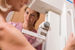
Women who undergo chest and abdominal CT exams may be at increased risk for breast cancer later in life, according to a study published September 14 in Applied Radiation and Isotopes.
The findings highlight the need to consider the effects of CT radiation dose on the breasts when women are undergoing the exam for reasons other than breast imaging, wrote a team led by Nissren Tamam, PhD, of Princess Nourah Bint Abdulrahman University in Riyadh, Saudi Arabia.
"Breast tissue is susceptible to radiation ... [and] breasts are constantly encompassed in the radiation primary field of chest CT even though it is not an organ of interest," the group noted.
Each year, 400 million CT procedures are performed, accounting for 62% of total annual collective effective dose for patients around the world, the investigators wrote. They further noted that chest CT exposes individuals to effective radiation doses 400 times those of x-rays.
This level of radiation exposure increases the risk of developing cancer, and breast cancer is definitely on the list: The team cited research that found breast tissue dose can vary four-fold depending on CT protocols and dose estimation practices -- thus boosting women's vulnerability to the disease.
"The high breast doses associated with CT imaging for the chest and abdomen imply that special attention is needed to optimize dose delivery for the maximum benefits of this undeniably beneficial diagnostic modality," Tamam's group wrote.
Tamam and colleagues conducted a study that included 28 women between the ages of 15 and 35 (average age, 25) who underwent chest and abdominal CT for other indications at three different hospitals in Saudi Arabia. No dose optimizations or reductions were made.
The group tracked dose length product (DLP, measured in mGy*cm) and CT dose index (CTDIvol, measured in mGy), effective dose (measured in mSv) and breast dose (measured in mSv), and found a wide range of discrepancies across the facilities, and determined that the average breast dose during CT chest was 10.2 mSv -- equivalent to three mammography exams.
| Radiation dose to women from chest and abdominal CT | ||
| Measure | Chest | Abdomen |
| CTDIvol | 9.1 to 36.2 mGy | 9.6 to 180.1 mGy |
| DLP | 474 to 1,506 (mGy*cm) | 524.6 to 1,282 (mGy*cm) |
| Effective dose | 6.6 to 21 (mSv) | 7.9 to 19 (mSv) |
| Breast dose | 4.6 to 15 (mSv) | 5.5 to 13.4 (mSv) |
Why the variation? It could have to do with training and the range of clinical indications -- and addressing the problem could mean optimizing protocols and making better use of dose-saving software, the group noted.
"Unoptimized protocols may also contribute to patients receiving more radiation exposure. In many hospitals, limited training on new technology affects the technologist's ability to properly use dose-saving software to reduce patient doses to possible values," the researchers noted.
What's the risk that radiation doses at these levels would result in cancer? The authors cited previous research indicating that the likelihood of cancer risks due to CT chest exams is 1.2 cases per 1,000 procedures and that the linear no-threshold theory maintains that cancer risk is most likely dependent on the level of radiation dose.
"Therefore, there is a need for dose reduction techniques in young females due to their higher risk of cancer," the researchers concluded.


