Cross-sectional supplemental imaging with MRI and CT can improve sensitivity when detecting tumors in axillary lymph nodes in breast cancer patients, a German study published August 10 in the Journal of Cancer Research and Clinical Oncology found.
Researchers led by Dr. Joachim Diessner from the Josef-Schneider-Strasse University Hospital in Würzburg found that using a combination of MRI and CT has a higher sensitivity than other combined imaging methods, allowing for safe detection of these lymph nodes at the time of diagnosis.
"Only the safe detection... enables the evaluation of the response to neoadjuvant therapy, thereby allowing access to prognosis and improving new post-neoadjuvant therapies," Diessner and colleagues wrote.
The status of axillary lymph nodes is one of the most important prognostic factors for determining long-term survival of breast cancer patients. This includes finding out whether the nodes are infiltrated by tumors, since nodal involvement influences treatment decisions.
Supplemental imaging is used to evaluate lymph node status. While a combined method using ultrasound and mammography has been a go-to choice for radiologists, its sensitivity has a wide range depending on what previous research is cited. MRI and CT have also been explored for such supplemental imaging. However, research shows that these have lower specificity than conventional approaches.
Diessner and colleagues wanted to explore the sensitivity of pretherapeutic imaging modalities in nodal-positive breast cancer patients. They used sonography, mammography, MRI, and CT imaging. The researchers wanted to find out what further benefits could be had with cross-sectional imaging using MRI and CT for axillary staging compared with the mammography and sonography combination.
The team looked at retrospective data from 382 women who received surgery between 2014 and 2020, including 201 women who received cross-sectional imaging.
| Sensitivity of combined imaging methods for axillary staging of lymph nodes in breast cancer patients | |
| All imaging modalities | 68.89% |
| MRI/CT | 63.83% |
| Mammography/sonography | 36.11% |
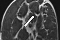
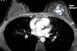
As an example, the researchers noted the results of a 39-year-old patient with hormone receptor-positive, HER2-negative breast cancer. They wrote that while CT at the initial staging of the thorax and abdomen gave suspicion of lymph node infiltration, conventional imaging couldn't clearly represent the tumor-infiltrated lymph node.
"During the period of data collection, we could detect an increasing importance of cross-sectional imaging," the authors wrote about the MRI and CT combination.
They added that they could not prove any effect on imaging sensitivity for lymph node status when accounting for other clinical parameters such as age, intrinsic subtype, histological subtype, and body mass index.
Diessner et al noted that while prospective data would be "highly interesting," such a study would be challenging when it comes to implementation and patient recruitment. Still, the team touted cross-sectional imaging with MRI and CT as being able to help guide treatment decisions with neoadjuvant chemotherapy with improved sensitivity.