Interpreting ultrasonic images for use in plastic surgery can be challenging, including when it comes to lymphatic vessels or when no other information is available, a Spanish study published on 28 June in the Journal of Plastic, Reconstructive & Aesthetic Surgery found.
Researchers led by Paloma Malagón, PhD, from Germans Trias i Pujol Hospital in Barcelona found that while plastic surgeons had lower accuracy than radiologists for interpreting ultrasound images, they gained on radiologists upon second assessment.
"To our knowledge, this is the first study that compares the cognitive ability of plastic surgeons to interpret the sonographic images with other medical specialists and that establishes an order of difficulty to recognize the main anatomical structures and layers for reconstructive surgeries," Malagón and colleagues wrote.
Ultrasound has become more popular in plastic surgery settings with previous research touting its noninvasiveness and high accuracy, and how it's well-tolerated by patients. However, specific training is needed for its use in this area. Plastic surgeons, like radiologists, must learn about ultrasound's manual ability, as well as how to acquire and interpret images.
Malagón and colleagues wanted to compare the ability of plastic surgeons with that of radiologists and medical students for identifying features in ultrasound images.
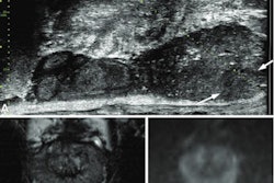
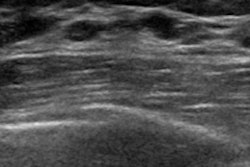
The team had a total of 30 observers identify 46 anatomical structures and layers included in 30 sonographic images. Ten observers were plastic surgeons, 10 were radiologists, and the remaining 10 were students. Images of the skin, subcutaneous tissue, muscle, and bone layers were included. While B-mode imaging was used for most ultrasound images included in the study, color Doppler and pulsed-wave Doppler were also used for a few images.
Participants had a maximum response time of 30 seconds. Researchers gave no answer options or other information except the word "compression" in the image where it was applied. A total of 1,380 answers were submitted.
The researchers also asked respondents to rate their confidence in their interpretation on a scale of 0 to 5, with higher scores indicating a greater level of confidence.
| Plastic surgeons vs. students and radiologists for interpreting ultrasound images | |||
| Students | Radiologists | Plastic surgeons | |
| Correct response rate | 31% | 76.5% | 69% |
| Confidence in responses | 1.7 | 4.1 | 2.5 |
The team also randomly showed images that included 10 layers and structures two times to each participant. The rate of correct responses was higher among the plastic surgeons, increasing from 73% in the first test to 87% in the retest (p = 0.04). The confidence level of plastic surgeons also received a boost upon retest, increasing from 2.4 to 2.7 (p = 0.037).
Plastic surgeons had an intraobserver agreement value of 0.79 compared with 0.87 for radiologists. The surgeons also had an intraobserver concordance index of 83% versus 90% for radiologists.
The researchers found that for study participants, lymphatic vessels, nerves, and veins were the most difficult images to identify. The authors wrote that for all three cohorts, correct response rates for anatomical layers were 76.8% and 50.7% for structures. However, they added the plastic surgeon cohort did best at identifying lymphatic vessels.
"These differences would be explained by their higher experience in ultrasound examination of lymphatic vessels compared with the radiologists, and their greater anatomical knowledge compared with the students," the researchers wrote.
Malagón and colleagues wrote that their results may suggest a learning process based on the improvement of the plastic surgeons on a retest.
"The first time an image is assessed, all the possibilities are considered, and the evaluation requires more time and effort," they added. "Once the decision is taken, it is less difficult to answer when the same image is evaluated again some minutes later."
Previous research also suggests that beginners can improve their ultrasound skills after attending learning sessions. The authors wrote this means the learning curve is "relatively short" for some assessments, such as identifying anatomical layers or superficial veins.