Danish nuclear medicine physicians recently tested a new whole-body PET/CT scanner with a five-minute image acquisition time and no sedation on a 17-month-old girl to resolve a case of suspected incomplete Kawasaki disease.
With the mother present and singing to the child to calm her, the girl was positioned arms-free in a vacuum fix pillow supplemented with light fixation across her body using a Velcro belt. Low-dose CT was followed by a five-minute PET acquisition in list mode, wrote a team led by Dr. Barbara Fischer, PhD, of Rigshospitalet in Copenhagen in a report published June 16 in the Journal of Nuclear Medicine.
"The images were of good quality for interpretation despite slight misalignment over the extremities," Fischer and colleagues noted. "PET/CT demonstrated no signs of infection or malignancy."
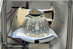
Whole-body PET and long axial field-of-view (LAFOV) PET are game-changing innovations at the threshold of clinical implementation, according to the authors. In this study, the physicians used a LAFOV PET/CT scanner (Biograph Vision Quadra, Siemens Healthineers) installed at Rigshospitalet in September 2021.
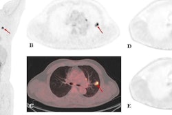
 CT, PET, and fused PET/CT in sagittal, axial and maximum intensity projection reconstructions (A-D) after 120 second PET acquisition. Color scale from 0-5 standard uptake value (SUV). No pathological uptake, but reactive accumulation in the distal part of esophagus (yellow arrow), physiological thymic uptake (blue arrow), and accumulated urinary activity in the diaper (white arrow).
Research suggests that these new scanners can help reduce the use of sedation in children by acquiring images faster, given that PET/CT acquisition times on many current systems in clinical use can take up to 20 minutes and motion affects the quality of imaging. The major advantage is the new system's 106-cm axial field of view, which is four times that of even the latest clinically established whole-body PET/CT scanners. Early experience shows high sensitivity, time-of-flight resolutions, and improved image quality, the authors wrote. But studies beyond research purposes are scarce, and initial clinical experiences are just beginning to be reported.
In this case, the girl was evaluated for incomplete Kawasaki disease on F-18 FDG whole-body PET/CT after 12 days of fever, despite treatment with broad-spectrum antibiotics. Previously, she had left heminephrectomy due to duplex kidney and repeated urinary tract infections. She had high C-reactive protein, anemia, hypoalbuminemia, and thrombocytosis, and her fever had relapsed despite immunoglobulin therapy and high doses of acetylsalicylic acid.
She underwent a PET/CT scan 74 minutes after receiving an injection of F-18 FDG radiotracer (3 megabecquerels/kilogram) that consisted of a low-dose CT followed by a five-minute PET acquisition. An image frame of 120 seconds with minimal movement was reconstructed using a standard protocol.
Results indicated no signs of infection or malignancy on the PET/CT scan, and the patient was discharged, with all parameters normalized at follow-up a week later, the authors wrote.
"This case illustrates how LAFOV PET enables whole-body PET imaging in children without the risks and logistical challenges associated with sedation," they concluded.