Data derived from MRI exams show that body composition can change significantly over a two-year period, potentially offering a new window into the aging process, according to research published recently in Scientific Reports.
A team of researchers from the U.K. led by Brandon Whitcher, PhD, of the University of Westminster in London studied over 3,000 patients in the UK Biobank who received knee-to-neck MRI scans at their first imaging visit and then again two years later. They found significant changes in visceral adipose tissue, intermuscular fat, and grip strength, even in participants without increased body mass index and waist size.
"The capacity to track quantitative changes in body composition of an individual, at the global and granular levels including multiorgan assessment, gives us a unique opportunity to objectively and quantitatively determine the impact of day-to-day factors on health and the aging process," co-author Jimmy Bell, PhD, of the University of Westminster told AuntMinnie.com.
In their study, the researchers ought to determine if longitudinal change in organ volume and composition would be detectable in a large population living in an obesogenic environment. Secondarily, they wanted to evaluate whether these changes would be accelerated or ameliorated in individuals living with a chronic disease or physiological condition.
In 3,088 cases from the UK Biobank, deep-learning algorithms were used to segment and perform volumetric assessment of the liver, pancreas, spleen, and kidneys on MRI. The researchers also evaluated for changes in adipose tissue, skeletal muscle, and the vascular system. What's more, they measured the levels of proton density fat fraction and iron concentration for each participant.
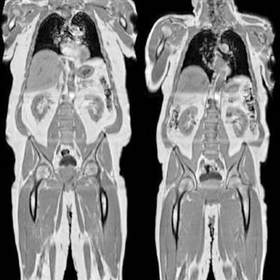
 Examples of change in body mass index for three UK Biobank subjects. From left to right: -6.9, 0.0, and 4.4 kg/m2. Images courtesy of Brandon Whitcher and the UK Biobank.
Using an automated image processing pipeline, 27 image-derived phenotypes were generated from the abdominal MR protocol and then combined with physiological measures related to the disease or physiological condition. After identifying combinations of image-derived phenotypes and disease, the researchers found that the trajectory of longitudinal changes in body composition image-derived phenotypes was dependent on the study participants' physiological state.
Although the authors didn't find any significant changes in body mass index, body weight, or waist circumference among the participants, they did observe significant increases in visceral adipose tissue and intermuscular fat in the thighs on the MRI exams, as well as a significant reduction in grip strength: a 2.3% decrease in women and a 4.3% decrease in men. What's more, they also spotted small but statistically significant decreases in all skeletal muscle measurements.
Even after a relatively short period of time, significant body composition changes can occur, probably reflecting the obesogenic environment currently inhabited by most of the general population in the U.K., the researchers concluded. These changes could have significant repercussions on muscle quality and function, and they may also serve as a precursor to subsequent health conditions, the study authors said.
"As larger datasets become available, we can start thinking about moving away from preclinical models to a point where human participants are the main source of information in the fight against obesity and accelerated aging," Bell said.