Shear-wave elastography used preoperatively can predict the success of rotator cuff surgery, according to research published January 20 in the American Journal of Roentgenology.
A team led by Dr. Jeung Yeol Jeong from Hallym University Medical Center in South Korea found that measurements of tissue elasticity taken from elastography scans were higher in patients with insufficient rotator cuff repair. The group also found that one metric, elasticity ratio, independently predicted insufficient repair.
"The findings indicate a complementary role for shear-wave elastography as a prognostic marker during preoperative evaluation in patients with rotator cuff tear," Jeong and colleagues wrote.
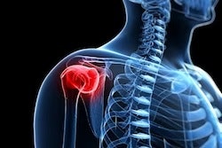
The prevalence of rotator cuff tears varies around the world, but research suggests that it is generally increasing. Such tears cause shoulder pain and dysfunction while the torn muscle shows fatty degeneration and atrophy linked to a loss of elasticity.
For successful repair, accurate evaluation of muscle quality before surgery is needed to guide prognostic assessments. MRI is one imaging method used to assess pathology and muscle quality. However, it does not give information on muscle tensile force or stiffness.
Shear-wave ultrasonography has shown promise in musculoskeletal imaging, such as evaluating pathologies of the supraspinatus tendon of the rotator cuff. "There have been few previous studies on supraspinatus muscles and a small population has been used, and it is known that normal supraspinatus muscle has higher shear-wave elastography values," said corresponding author Dr. Eun Kyung Khil from Hallym University Dongtan Sacred Heart Hospital. Jeung et al wanted to look at the use of preoperative elastography measurements to predict successful rotator cuff repair and compare them with MRI-based measures.
They looked at data from 74 patients with an average age of 63.9 years. Out of these, 37 were men and 37 were women. Study participants underwent rotator cuff repair between May 2019 and January 2021.
Patients underwent preoperative MRI and shear-wave elastography, with the average elasticity measured for the supraspinatus and trapezius muscles. The elasticity ratio between supraspinatus and trapezius mean elasticity was also calculated.
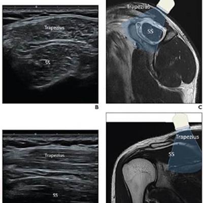
 (A) A 37-year-old male patient is in a supine and neutral position, with his neck muscle gently stretched and head turned slightly to the contralateral side. The ultrasound transducer is initially placed with application of minimal pressure. (B) The supraspinatus and trapezius muscles are identified in the transverse plane. (C) The view is represented using a sagittal T2-weighted MR image. (D) The transducer is turned to the longitudinal view. (E) Supraspinatus and trapezius muscles are visualized along longitudinal orientation of muscle fibers. (F) View is represented using a coronal T2-weighted MR image. SS = supraspinatus
After surgery, 60 cases were determined to have sufficient repair while 14 were determined to be insufficient. Researchers found that patients who were deemed to have insufficient repair had a higher prevalence of large tears between three and five centimeters than patients who had sufficient repair (100% vs. 50%).
Jeung et al used different scales for each method. These include muscular fatty infiltration score (scale of 1 to 3), which was recorded on grayscale ultrasound; Goutallier's grade for muscular fatty infiltration measured by MRI (scale of 0 to 4); and muscular atrophy assessed by the occupation ratio and by muscle atrophy grade (scale of 0 to 3).
| Performance of MRI, shear-wave elastography measurements for predicting rotator cuff repair success | ||||||
| MRI | Shear-wave elastography | |||||
| Muscle atrophy grade | Goutallier's grade | Occupation ratio | Mean elasticity (kPa) | Grayscale fatty infiltration grade | Elasticity ratio | |
| AUC | 0.900 | 0.945 | 0.961 | 0.874 | 0.912 | 0.971 |
| Sensitivity | 85.7% | 78.6% | 85.7% | 92.9% | 85.7% | 100% |
| Specificity | 90% | 95% | 96.7% | 70% | 90% | 90% |
After analysis of tear size, the three MRI measures, elasticity ratio, and grayscale fatty infiltration grade, the study authors wrote that the only independent predictors of insufficient repair were muscle atrophy grades of 2 to 3 (odds ratio, 9.3) and elasticity ratio (odds ratio, 15.6).
For elastography measures, patients who had insufficient rotator cuff repair had higher mean supraspinatus elasticity, elasticity ratio, and grayscale fatty infiltration grades than patients who had sufficient repair.
Jeung and colleagues wrote that these findings support a potential role for shear-wave elastography in preoperatively assessing rotator cuff repair success, with the emergence of ultrasound elastography allowing for tissue evaluation by elasticity measurements.
"We believe that our study makes a significant contribution to the literature because we found that shear-wave elastography values of the supraspinatus muscle exhibit a positive correlation with the degree of muscle quality, which would be useful in predicting tendon reparability during surgical planning," Khil told AuntMinnie.com.