The combination of CT radiomics and machine learning can predict survival of melanoma patients after immunotherapy better than relying only on tumor size data, according to research published online January 20 in JAMA Oncology.
A multi-institutional team of researchers led by Dr. Laurent Dercle, PhD, of Columbia University Medical Center developed a machine-learning algorithm and a radiomics "signature" for estimating overall survival from CT images in patients receiving immunotherapy in two multicenter immunotherapy clinical trials. In testing, the radiomics signature outperformed the Response Evaluation Criteria in Solid Tumors (RECIST) 1.1 guideline in predicting overall survival at six months.
"The findings of this prognostic study suggest that the radiomic signature discerned from conventional CT images at baseline and on first follow-up may be used in clinical settings to provide an accurate early readout of future [overall survival] probability in patients with melanoma treated with single-agent programmed cell death 1 blockade," the authors wrote.
Although existing criteria rely almost exclusively on tumor size to estimate therapy benefit in cancer patients, this approach wasn't designed to estimate survival benefit and is also challenged by the unique properties of immunotherapy, according to the researchers
"With a principal goal of improving clinical management, we tested the hypothesis that a radiomic signature derived from tumor size, density, and shape and its change as treatment is administered can estimate [overall survival] in patients with melanoma," the authors wrote.
They utilized data provided by the Advanced Metrics and Modeling with Volumetric CT for Precision Analysis of Clinical Trial Results (Vol-PACT) public-private partnership to retrospectively analyze CT scans and clinical data from two landmark randomized clinical trials for pembrolizumab. The analysis included 575 adults with unresectable stage III/IV melanoma.
After assessing 25 imaging features extracted from tumors segmented on CT images, a random-forest machine-learning model identified a radiomics signature that best estimates overall survival. This signature included two imaging features related to tumor size and two that reflect changes in tumor imaging phenotype.
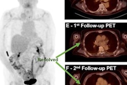
The researchers then assessed the final radiomics machine-learning model for assessing six-month survival on a CT scan performed three months after treatment on a validation test set of 287 patients treated with pembrolizumab.
| Ability of CT radiomics signature to predict survival of melanoma patients | ||
| RECIST 1.1 | Radiomics machine-learning model | |
| Area under the curve | 0.80 | 0.92 |
"This ability to discriminate among patients with progressive disease at month 3 could allow physicians to discuss alternative treatments earlier, appropriately set goals of care, or look for clinical trials," the authors wrote.
Because it only requires identification and segmentation of target lesions on routine CT scans, the radiomics and machine-learning prognostic model could be widely translated into clinical practice using publicly available software and a clinical decision-support software tool, according to the researchers.
They noted, however, that clinical application would currently be limited by the need for manual lesion segmentation, which requires approximately one minute per lesion per scan. However, deep-learning methods for automatic lesion detection and segmentation are being developed that would enable this task to be automatically performed after CT scans are acquired.
In an accompanying commentary, Dr. Michael Farwell and Dr. David Mankoff, PhD, of the University of Pennsylvania said that the trial represents an exciting milestone in the application of radiomics to routine CT scans from multicenter clinical trials.
"Although there are a few hurdles to overcome before this approach becomes part of routine clinical practice, it is only a matter of time before those tools are developed," Farwell and Mankoff wrote. "As a first step, tools that enable automatic lesion segmentation and change in volume would be a welcome addition to radiology clinical practice. Once these tools are established, it will be fairly straightforward to add radiomics to the analysis with improved prediction of survival for patients with cancer who are treated with immune checkpoint blockade."