Imaging findings on chest CT and x-ray in patients with breakthrough COVID-19 disease tend to be mild -- and most patients go on to recover just fine, according to research published December 9 in Radiology: Cardiothoracic Imaging.
"The majority of patients with breakthrough COVID-19 illness in this study had absent or mild imaging findings and a benign clinical course," wrote a group led by Dr. Rydhwana Hossain of the University of Maryland School of Medicine in Baltimore.
Vaccines to prevent COVID-19 appear to be effective, but some people still develop the disease -- no vaccine works 100%, the team wrote. Yet rates of these breakthrough infections appear to be low: A study conducted in Israel and published in July in the New England Journal of Medicine found that of 11,500 fully vaccinated people, only 0.34% developed breakthrough COVID-19, and all of these patients had mild or no symptoms.
Clinical aspects of breakthrough COVID-19 have been described, but imaging findings in these cases have yet to be thoroughly explored. Hossain's group sought to characterize chest x-ray and CT results in patients with breakthrough COVID-19 in a hospital setting. The team's study included eight patients out of 60 admitted to the hospital for COVID-19 symptoms between August and September; these eight were both fully vaccinated and had a positive reverse transcription polymerase chain reaction (RT-PCR) test for SARS-CoV-2. All patients had either chest x-ray, chest CT, or both.
Of the eight individuals, 63% were immunosuppressed when they were admitted, and 75% reported respiratory symptoms. Most (57%) had normal chest x-ray exams. On chest x-ray, common findings were hazy opacities and consolidation, while on chest CT, common findings were ground-glass opacities with mild to moderate severity scores. Two of the eight patients required admission to the intensive care unit. At the end of the study period, 75% were discharged (average length of stay: 13 days).
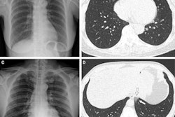
 (A) Axial chest CT on lung windows of a patient at presentation demonstrates bilateral right greater than left lower lobe consolidations with surrounding ground-glass opacities. (B) Follow-up image two weeks later demonstrates substantial improvement in bilateral lower lobe consolidations with minimal residual opacities and tiny effusions. Image and caption courtesy of the RSNA.
"Our results are in line with the clinical findings of COVID-19 breakthrough as reported in the literature, demonstrating that imaging findings in such patients are commonly mild ... and outcomes generally favorable," Hossain and colleagues wrote.
They did acknowledge that one of the study's limitations is its small sample size, and they urged continued tracking of imaging findings as clinicians encounter more breakthrough COVID-19 cases.
"As the number of COVID-19 breakthrough cases is likely to increase, it will be important to continually document imaging findings to determine if the imaging patterns remain consistent with those observed in this study or whether they evolve," the authors concluded.