Diffusion-weighted imaging has made strides in research on its way to being used in radiology clinics for diagnostic and therapeutic use in breast imaging, but more work is needed, according to a talk given December 1 at the RSNA annual meeting.
In her keynote presentation, Savannah Partridge, PhD, from the University of Washington touted this imaging method's potential for classifying breast lesions, evaluating response to therapy, and not using contrast agents to detect cancers.
"Diffusion-weighted imaging can generate a wealth of information related to tissue microstructure," Partridge said. "It's already shown clinical value for multiple applications for breast imaging."
Diffusion-weighted imaging uses diffusion-sensitizing gradients during image acquisition for radiologists to measure water motion. For breast imaging, malignancies show restricted diffusion on diffusion-weighted imaging compared with normal breast tissue, presenting as bright spots.
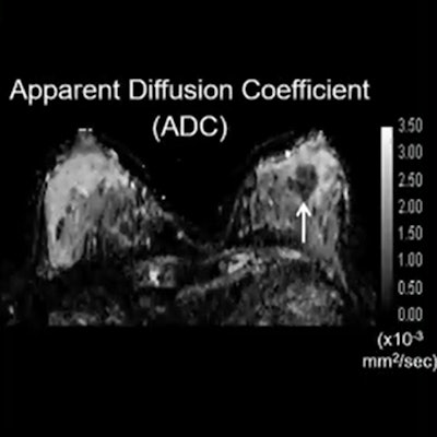
 Malignancies present as bright spots on diffusion-weighted imaging due to showing restricted diffusion. Researchers say this imaging method could serve as an alternative to dynamic contrast-enhanced MRI, though more studies and standardization are needed. Image courtesy of Dr. Savannah Partridge.
"It's this common characteristic of malignancies that make it [imaging] value for breast imaging," Partridge said.
While progress has been made in this technology over the past 20 years, it has not been established as a routine clinical technique.
Previous studies, one of which Partridge led in 2018, have shown that malignancies detected by diffusion-weighted imaging have lower apparent diffusion coefficient (ADC) values than benign lesions. Partridge and colleagues also suggested that ADC could improve specificity and positive predictive value of conventional breast MRI.
Studies have also shown diffusion-weighted imaging's promise in evaluating response to neoadjuvant therapy by showing alterations in cell membrane integrity and reduced cell density, leading to increased water mobility in the tumor microenvironment.
Partridge led another study in 2018 showing that midtreatment ADC was more predictive of pathologic complete response than changes in tumor size shown by dynamic contrast-enhanced MRI. Tumor ADC has also shown repeatability in test-retest scenarios.
Partridge also noted diffusion-weighted imaging's potential use as an alternative noncontrast screening method. One study Partridge cited from 2019 that compared diffusion-weighted imaging with dynamic contrast-enhanced MRI showed the former had an average sensitivity of 72% and a specificity of 90%.
"This is not yet reaching the performance of dynamic contrast-enhanced performance, but it's quite promising for a technique that has not been fully optimized for this application," Partridge said.
One current research effort Partridge noted is being performed by South Korean researchers. The team is enrolling women into the prospective multicenter trial, which aims to compare diffusion-weighted imaging with other breast screening methods.
"We really look forward to their results to help shed more light on the value of diffusion-weighted imaging for this application," Partridge said.
However, one barrier preventing this imaging method from being used routinely in clinics is the lack of standardization. This has limited researchers in terms of defining diagnostic criteria and integrating diffusion-weighted imaging into BI-RADS and other guidelines.
However, efforts are being made by groups in Europe and the U.S., including the EUSOBI International Breast Diffusion-Weighted Imaging working group and the RSNA's Quantitative Imaging Biomarkers Alliance.
Partridge also said more multicenter studies are needed to further validate this method and evaluate its real-world applications, with emerging image acquisition techniques seeking to expand its utility and role in breast imaging.
"There's still exciting things to come," she said.