Chest x-rays performed three months after patients with COVID-19 are discharged from hospital can identify those who might need additional follow-up treatment, according to a study published October 22 in Radiography.
Italian researchers analyzed chest x-rays of COVID-19 patients acquired at admission, prior to discharge, and three months later and found abnormalities were present in about half of patients. Based on a scoring system the authors used, the finding could help prioritize patients with more severe clinical and radiological findings, the authors wrote.
"The radiologist with [chest x-ray] could play a central role in mid- to long-term follow-up of COVID-19, assessing the radiological sequelae of patients and identifying those who might require a closer follow-up," wrote lead author Dr. Marco Fogante of Azienda Ospedaliera Ospedali Riuniti in Ancona, Italy.
Studies on mid- or long-term radiological follow-up of the lasting effects of COVID-19 in patients are scarce, according to the researchers. Follow-up x-ray exams could help evaluate the development of irreversible fibrosis in these patients, for instance. However, the optimal time for follow-up imaging is unknown, the authors wrote.
In this study, the Italian team sought to evaluate radiological sequelae of COVID-19 in patients at three months and investigate their relationship with clinical-radiological findings.
From May to June, 2021, the researchers enrolled 119 consecutive patients with a mean age of 66 years old (+/- 14.6 years) for a follow-up chest x-ray three months after being discharged. All patients had received chest x-rays at admission and before discharge and had COVID-19 confirmed by a lab test.
Two radiologists evaluated the chest x-rays of patients at admission, at discharge, and at three months using an 18-point scoring system. The scoring system divided lungs into three equal parts: upper, middle, and lower, for a total of six zones. Scores from 0 to 3 were assigned to each zone based on lung abnormalities detected on a frontal view, as follows:
- 0 -- No abnormalities
- 1 -- Interstitial infiltrates; defined as septal thickenings and focal or extensive opacity, with the evidence of extravascular structure
- 2 -- Interstitial and alveolar infiltrates (interstitial predominance)
- 3 -- Interstitial and alveolar infiltrates (alveolar predominance)
The single scores of the six lung zones in each patient were added to obtain an overall chest x-ray score ranging from 0 to 18. Patients were divided into groups with scores of 0 or scores ≥ 1, and clinical-radiological findings were compared between them.
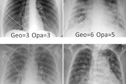
 An example of a patient with a chest x-ray score of 6 at admission and scores of 4 before discharge and at three months follow-up. Image courtesy of Radiology.
About half of patients (49.6%) had chest x-ray scores of 0, which indicated a complete radiological recovery three months after discharge. The other half (50.4%) had x-ray scores of 3 (+/- 2.6) at three months. In patients with higher scores at three months, age, the number of days of hospitalization and their chest x-ray scores at admission and before discharge were also statistically higher, the researchers found.
"Radiological abnormalities persist three months after discharge in a high proportion of COVID-19 patients," the authors wrote.
Ultimately, the study suggests that evaluating chest x-rays scores at admission and before COVID-19 patients are discharged can identify those who may benefit from a closer follow-up and targeted management, the researchers concluded.