CT radiomics and a machine-learning model can predict hematoma expansion in patients with spontaneous intracerebral hemorrhage (ICH), according to a presentation at the recent Conference on Machine Intelligence in Medical Imaging.
After experimenting with a variety of different machine-learning models, researchers from China found that a model that analyzed both CT radiomics and clinical features yielded an area under the curve of 0.9 on a set of test cases.
"The study showed the feasibility of radiomics in predicting hematoma expansion in spontaneous intracerebral hemorrhage, which can help clinical intervention and [enable] personalized medicine," said presenter Zekun Jiang of Sichuan University's West China Biomedical Big Data Center.
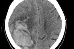
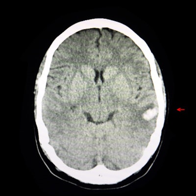
Spontaneous ICH is a dangerous clinical condition that has high disability and mortality rates, and hematoma expansion is associated with poor prognosis in these patients.
"Early identification ... can lead to preventive interventions, helping [to] improve prognosis," Jiang said.
As a result, the researchers sought to create an optimal CT-based radiomics model for predicting expansion of hematoma in patients with spontaneous ICH. They first retrospectively gathered data from 235 patients at West China Hospital who had spontaneous ICH. Of these patients, 80 (34%) had hematoma expansion.
All patients in the study received noncontrast CT within six hours after symptom onset and then follow-up CT within 24 hours after onset. The researchers used 75% of the exams to train the model and set aside 25% for testing.
Segmentation of regions of interest on the CT exams was performed on the ITK-Snap 3D image segmentation software. Radiomics features -- including 1,106 first-order, texture, shape, and wavelet features -- were then extracted with the open-source pyradiomics software.
Next, the researchers constructed nine different machine-learning models using one of three radiomics feature selection methods -- least absolute shrinkage and selection operator (LASSO), random forest, or support vector machine (SVM) -- recursive feature elimination -- and one of three classifiers: logistic regression, random forest, or SVM.
The best-performing model used LASSO for feature selection followed by logistic regression for classification. LASSO selected seven optimal radiomics features for analysis, including six texture features.
This "fully showed the heterogeneity between the lesions," Jiang said.
Four clinical features -- initial hematoma volume, diastolic blood pressure, satellite sign, and blend sign -- were significantly associated with hematoma expansion (p < 0.05).
In testing, the researchers found that a combined radiomics-clinical model outperformed both a radiomics model and a clinical model.
| Performance for predicting hematoma expansion in spontaneous ICH | |||
| Clinical model | Radiomics model | Radiomics-clinical model | |
| Area under the curve | 0.80 | 0.85 | 0.90 |
"The radiomics-clinical model constructed by [logistical] regression after LASSO selection was the best predictive model, which may help early diagnosis [of spontaneous ICH] hematoma expansion," Jiang said.
The researchers acknowledged the limitations of their study, including its single-center, retrospective nature and the imbalance between cases with and without hematoma expansion. In addition, randomized datasets are biased, and should be divided according to imaging time or scanner type, he said.
"This is a preliminary exploratory study and needs to be validated with large, multicenter data," Jiang said.