PET imaging has shown that an experimental radiotracer can detect cellular activity in the artery walls of patients with atherosclerosis, according to a study published September 18 in the Journal of Nuclear Medicine.
A Chinese team analyzed uptake of gallium-68 (Ga-68)-labeled fibroblast activation protein inhibitor (FAPI) in the arteries of patients with suspected atherosclerosis. They found uptake of the tracer was more intense in the arteries of patients with less plaque than in those with severe lesions -- a finding that suggests Ga-68 FAPI-PET/CT can identify early activity involved in the progression of the disease.
"Our preliminary study provides a potentially feasible method to image atherosclerosis in vivo by Ga-68 FAPI-PET/CT," wrote first author Dr. Meiqi Wu of the department of nuclear medicine at Peking Union Medical College Hospital, China, and colleagues.
The fatty build-up of plaque in the arteries (calcification) is a hallmark of atherosclerosis. Researchers suspect inflammation caused by an enzyme known as fibroblast activation protein (FAP) is involved in causing plaque to form.
Preliminary Ga-68 FAPI-PET/CT studies have shown promising results in detecting fibroblast activation protein activity in cancer and lung fibrosis. Recently, however, the tracer has also shown in animal and lab studies that it also may be effective in assessing fibroblast activity in atherosclerosis.
In this study, Wu and colleagues aimed to quantify arterial fibroblast activation with Ga-68 FAPI in human patients for the first time and determine whether it is associated with known risk factors for atherosclerosis.
The researchers analyzed data from 41 patients (10 women and 31 men) who underwent Ga-68 FAPI-PET/CT on a dedicated hybrid scanner (Polestar m660, SinoUnion, China) for noncardiovascular indications at Peking Union Medical College Hospital between January 2019 and January 20. They measured maximum standardized uptake values (SUVmax) and target-to-background ratios (TBR) in noncalcified, mildly calcified, and severely calcified artery segments.
In addition, patients were divided into high-risk (prevalence of ≥ 4 cardiovascular risk factors) and low-risk (< 4 cardiovascular risk factors) groups.
The researchers identified a total of 1,177 arterial segments of focal uptake of Ga-68 FAPI or calcified plaque in all 41 patients. Among all of the assessed arterial segments, the abdominal aorta exhibited the highest number of segments (n = 379), followed by the thoracic aorta (n = 272) and the ascending aorta (n = 203).
The correlation between the extent of plaque and the intensity of Ga-68 FAPI uptake was significant in all 1,177 artery segments. Noncalcified segments presented with significantly higher uptake (TBR = 2.2; n = 119) than mildly calcified segments (TBR = 1.9; n = 220), while severely calcified segments exhibited the lowest uptake of Ga-68 FAPI, the researchers found.
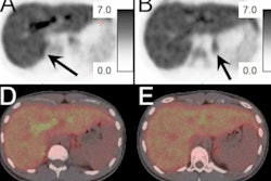
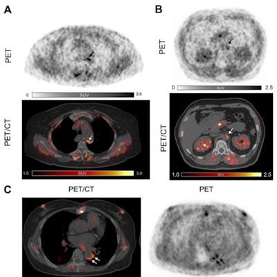
 Three examples of Ga-68 FAPI uptake of active arterial segments. All three patients were over 60 years old with a history of hypertension and dyslipidemia. Patients A and C also had diabetes mellitus, and experienced myocardial infarction and percutaneous coronary intervention treatment. Patient A was obese (body mass index = 30) while patient B had a history of heavy smoking.
"To our knowledge, this is the first noninvasive study to describe the expression of FAP in the human arterial walls via Ga-68 FAPI-PET/CT imaging," they wrote.
In addition, the researchers found obese patients specifically had more prominent arterial tracer uptake compared with patients with other cardiovascular risk factors.
"We observed elevated Ga-68 FAPI uptake in patients with increased cardiovascular risk factors," they stated.
The authors noted limits to the study, namely that it included patients who underwent imaging for noncardiovascular indications, such as suspicious liver lesions and immunoglobulin G4-related disease and that scans were performed using a routine protocol that was not optimal for vessel imaging.
Nonetheless, overall, the study provides a potentially feasible method to image atherosclerosis in patients in clinical settings by Ga-68 FAPI-PET/CT, the authors concluded.
"Prospective studies using Ga-68 FAPI-PET imaging in symptomatic atherosclerotic cohorts are warranted," they wrote.