Ultrahigh-resolution CT (UHR-CT) shows promise for overcoming conventional CT's limitations when it comes to imaging heart stenosis in patients with severely calcified lesions, according to a study published August 26 in Radiology: Cardiothoracic Imaging.
The findings could lead to more effective use of the modality for this application, wrote a team led by Dr. Jacqueline Latina of Johns Hopkins University.
"This study's initial experience with coronary angiography using ultrahigh-resolution CT in patients suspected of having coronary stenoses demonstrates promise for high diagnostic accuracy even in the setting of severe coronary calcification or prior stenting, representing a potentially substantial advance compared with conventional CT technology in this challenging patient population," the group noted.
Coronary CT angiography (CTA) has become the go-to modality for diagnosing patients with suspected coronary heart disease. But conventional CT tends to have only moderate accuracy when assessing coronary artery stenosis in severely calcified lesions, according to the researchers. What mitigates this limitation is higher spatial resolution, and UHR-CT offers this, they wrote.
Latina's team investigated the performance of ultrahigh-resolution CT with a commercially available scanner (Aquilion Precision, Canon Medical Systems) in patients with severe artery calcification. The study included 15 patients ages 45 or older with a history of coronary artery disease who had been referred for coronary angiography.
Each patient underwent ultrahigh-resolution CT within 30 days before cardiac catheterization; the researchers compared image noise levels and diagnostic confidence and image quality (the latter two measures ranked on a scale of 1 to 5, with 5 indicating highest quality) from these images to reconstructed images simulating conventional CT exams. They also compared UHR-CT and angiography performance for assessing stenosis in the major heart arteries and the left main artery.
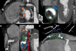
 Example of a 61-year-old man with a history of myocardial infarction who was found at UHR-CT to have a severe (≥ 70%) stenosis in a large obtuse marginal branch of the left circumflex artery just distal to (A) a patent stent, which was confirmed at (B) invasive angiography. In addition, there was moderate disease noted by (C) ultrahigh-resolution CT in the left main and in the middle left anterior descending artery (50%-70% stenosis), which was underestimated compared with (D) invasive angiography (70% stenosis). Arrow points to corresponding stenoses. Images and caption courtesy of the RSNA.
UHR-CT produced more image noise than conventional CT image reconstruction (50.9 versus 19.5), but even so, diagnostic confidence and image quality scores were high (4.3 and 4.1), the researchers found. When they compared the performance of ultrahigh-resolution CT to the gold standard of invasive angiography for evaluating stenosis, they found a sensitivity rate of 86% and specificity of 88%.
The study presents "preliminary evidence that ultrahigh-resolution techniques may fulfill current unmet needs in diagnostic accuracy facing conventional CT technology," according to an accompanying editorial written by Drs. Sujata Shanbhag and Marcus Chen, both of the U.S. National Institutes of Health.
"With future refinements in UHR-CT technology, there is promise for improved patient risk stratification, plaque characterization, function flow reserve assessment, and microvascular disease assessment," Shanbhag and Chen wrote. "Technological advancements may not only lead to improvements in the use of coronary CTA for disease detection but may also play a role in tracking progression or regression of disease as a useful surrogate target for treatment response."