"Live long and prosper," as Mr. Spock from "Star Trek" might say -- in this case, to the musculoskeletal radiologists who found the character's Vulcan salute on x-ray can help diagnose a type of painful foot neuroma. Their findings were published July 14 in Skeletal Radiology.
"The Vulcan salute sign on conventional radiographs is specific for Morton's neuroma," wrote radiologist Dr. Julien Galley of the University of Zurich and colleagues.
Morton's neuroma was first described by Thomas Morton in the mid-1800s. It is a painful condition that affects the balls of the feet, most commonly the area between the third and fourth toes. The condition occurs when the nerve between the toe bones becomes inflamed. Women are about eight to 10 times more likely than men to develop the condition, likely because of shoe styles, the authors wrote.
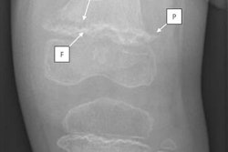
Morton's neuroma can lead to a divergence of the toes, which looks like the Vulcan salute on x-ray, according to the authors.
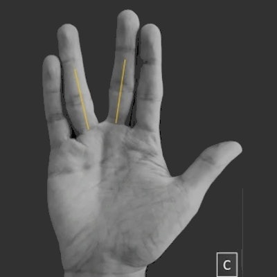
 (A) Negative Vulcan salute sign in a 50-year-old woman from the control group. (B) Positive Vulcan salute sign (V-sign) of the 3/4 interspace in a 46-year-old woman with Morton's neuroma. (C) "Vulcan salute" from one of the authors. Image courtesy of Skeletal Radiology.
For those unfamiliar with the "Star Trek" television series and the Mr. Spock character, the Vulcan salute (shortened to "V-sign" in the article) is a hand gesture made by parting the middle and ring fingers while the palm is facing forward and the thumb is extended.
In this retrospective case-control study, Galley and colleagues enrolled 100 patients with MRI-proven Morton's neuroma 2/3 or 3/4 (between the second and third or third and fourth toes) and 100 patients without the condition. Conventional weight-bearing dorsoplantar view radiographs were evaluated for the subjective presence of the V-sign by two blinded, independent musculoskeletal radiologists.
Conventional x-rays of the foot were taken from the standard weight-bearing dorsalplantar view, with the central ray directed over the middle of the third metatarsal. The x-ray tube was angled cranially at 15°.
The researchers found a significant difference between the groups regarding the presence of the V-sign, which was found in 30 of 100 patients in the Morton's neuroma group and in three of 100 control patients, with a sensitivity of 30% and a specificity of 97%.
The differences between interphalangeal angles were also significant between the groups, with interphalangeal angle 2/3 and 3/4 values higher for the study group than for the control group. Interobserver agreement was substantial for the V-sign, with a kappa value of 0.78.
While there was no discussion among the group regarding who is the bigger "Trekkie" ("Star Trek" fan), the researchers noted the V-sign on x-ray may not exclude Morton's neuroma, given its low sensitivity. Moreover, all of the patients included in the study underwent MRI scans for clinical indications and the exclusion criteria were broad, which could suggest selection bias.
Nonetheless, conventional radiography is recommended as the first radiological procedure by the American College of Radiology Appropriateness Criteria for the evaluation of chronic foot pain, and in cases of Morton's neuroma, x-rays may be helpful, the authors wrote.
"The presence of a V-sign on a plain radiograph is highly suggestive for the presence of a Morton neuroma," Galley et al concluded.